Neuron-Glia Crosstalk Underlies Senescence, Impaired Lipid Metabolism
Quick Links
Senescence, a state in which cells neither divide nor die, but simply linger, has been linked to aging and neurodegeneration. What causes such cells to appear in old brains, and how might they wreak havoc? In the June 5 Nature, researchers led by Nancy Bonini and China Byrns at the University of Pennsylvania, Philadelphia, offered clues gleaned from flies. They found that waning mitochondrial function in neurons triggered glial senescence. In turn, these senescent glia perturbed lipid metabolism in neighboring, healthy glia, which then accumulated lipid droplets. These oily droplets came with pros and cons. When the authors blocked their formation by preventing senescence, flies lived longer and were healthier, but their brains were more susceptible to oxidative damage. Going upstream and preventing mitochondrial damage might be a more effective way to bolster brain health, the authors suggested.
- Flagging mitochondria in aging fruit fly neurons push glia into senescence.
- Senescent glia promote lipid droplets in nearby, healthy glia.
- Reining in senescence extended lifespan, but at the expense of oxidative stress.
“This is exciting work that helps tie together several independent areas of research including cellular senescence, lipid droplet formation, and mitochondrial dysfunction during aging,” Hugo Bellen, Matthew Moulton, and Lindsey Goodman at Baylor College of Medicine in Houston wrote to Alzforum. Because mitochondrial dysfunction produces reactive oxygen species, antioxidant treatment might be an effective intervention to maintain the health of aging brains, they suggested (comment below).
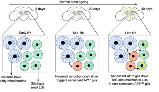
Cellular Crosstalk. In young flies (left), neurons (blue) and glia (green) are healthy, but in midlife (middle), mitochondrial damage in neurons triggers senescence in some glia (red). By late life (right), senescent glia drive lipid droplet (yellow) accumulation in healthy glia. [Courtesy of Byrns et al., Nature.]
Senescent cells appear in the mammalian brain with age, but had not been previously described in fly brain (Ito and Igaki, 2016). Byrns and Bonini earlier reported the first evidence of these cells, finding that fly glia switched on activator protein 1 (AP1), a transcription factor associated with senescence, during aging and after traumatic brain injury (Byrns et al., 2021).
To learn more about AP1-positive glia, and what they do, first author Byrns made use of a transgenic reporter line of flies that produce the fluorescent protein dsRed in cells in which AP1 is active. In young, 10-day-old reporter flies, dsRed was absent from the brain, but it appeared in glia in the antennal lobes in 20-day-old midlife flies, and in the optic lobe in late life, i.e., 40 days old.
Were these bona fide senescent cells? Byrns and colleagues isolated them and tallied their transcriptomes. This confirmed that dsRed glia had all the expected features: They were abnormally large, harbored damaged DNA, had heightened expression of metabolic and senescence-associated genes, and dampened expression of genes associated with cell division.
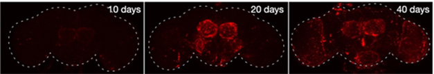
AP1 Marks Aging. Young flies (left) have no senescent cells (red) in their brains, but these gradually accumulate in mid- (middle) and late life (right). [Courtesy of Byrns et al., Nature.]
Because these senescent cells appear in the antennal and optic lobes around the time neurons begin to naturally die off there, Byrns wondered if neurodegeneration played a role, and compared neuronal gene expression in young and old flies. Older neurons produced less ATP and weakly expressed mitochondrial genes, suggesting problems with their energy supply. The authors tested this idea by using RNA interference to individually suppress genes important for these power plants, such as PINK1, OPA1, MARF, COX5A, NP15.6, ND42, and ND75. Lo and behold, each of these knockdowns made senescent glia appear in young, 10-day-old brains.
How might neuronal energy loss harm glia? Malfunctioning mitochondria are known to spark the formation of reactive oxygen species. Indeed, feeding an antioxidant to dsRed transgenic flies with damaged mitochondria prevented glial senescence. All told, the data hint that aging neurons could tip glia into senescence via oxidative stress.
Do these senescent glia affect the brain? RNA-Seq of senescent cells revealed they expressed more lipogenesis genes than did healthy glia. Co-author Gaurav Chopra at Purdue University, West Lafayette, Indiana, analyzed the cells’ lipidomes and found they accumulated about twice as much free fatty acid as did non-senescent glia. Free fatty acids can be converted to triacylglycerides, which make up lipid droplets. However, senescent cells did not form LDs; rather, these appeared in non-senescent glia, suggesting the senescent cells might be dumping lipids off to healthier cells.
In keeping with this, when the authors induced senescence in primary human fibroblasts for 10 days, then collected the media from these cultures and added it to fresh cells, those fibroblasts made lipid droplets. Not only does this suggest that secreted factors are responsible, it shows that mammalian cells can do the same, Bonini noted.
On whether blocking senescence would benefit the brain, the data were mixed. Suppressing AP1 activity for one day each week in adult flies indeed prevented lipid metabolism from going haywire, and kept LD content low. It also improved the flies’ lifespans, their climbing skills, and their ability to withstand heat, a measure of stress resistance in this species. However, flies missing AP1 altogether accumulated more oxidative damage in their brains when fed hydrogen peroxide, and, under that duress, died sooner than did controls. In addition, blocking senescence did nothing to improve neuronal mitochondrial health in normal flies.
In ongoing work, Bonini is crossing the dsRed reporter flies to models of neurodegeneration to parse what role senescent cells might play in these age-related diseases. Previous work has tied lipid dysregulation to neurodegeneration, with Chopra and Tony Wyss-Coray at Stanford University independently reporting that Aβ fibrils induce lipid droplets in microglia (Aug 2019 news; Sep 2023 news; Mar 2024 news). Chopra believes biological pathways to develop these droplets are conserved across species. “These senescent glia contributed to lipid droplet accumulation in non-senescent glia by similar lipid-related mechanisms implicated in age-onset diseases, such as Alzheimer's. This suggests that mitigating senescent glia may improve health, and opens up potential avenues for targeting aging and its diseases,” he wrote to Alzforum.—Madolyn Bowman Rogers
References
News Citations
- Newly Identified Microglia Contain Lipid Droplets, Harm Brain
- Lipid-Laden, Sluggish Microglia? Blame Aβ.
- Paper Alert: APOE4 Packs on Lipid Droplets in Microglia
Paper Citations
- Ito T, Igaki T. Dissecting cellular senescence and SASP in Drosophila. Inflamm Regen. 2016;36:25. Epub 2016 Dec 5 PubMed.
- Byrns CN, Saikumar J, Bonini NM. Glial AP1 is activated with aging and accelerated by traumatic brain injury. Nat Aging. 2021 Jul;1(7):585-597. Epub 2021 Jul 8 PubMed.
Further Reading
News
- Stirred by Tau, Neurons Amp Up Lipid Droplets in Glia
- While a Fly Sleeps, Its Glia Burn Neuronal Lipids to Refresh the Brain
- Paper Alert: APOE4 Packs on Lipid Droplets in Microglia
- Two Paths for TREM2-Positive Microglia: DAM or Senescence?
- When Autophagy Stops, Microglia Sour into Senescence
- DAMned to Death? Microglia May Proliferate to Senescence
Primary Papers
- Byrns CN, Perlegos AE, Miller KN, Jin Z, Carranza FR, Manchandra P, Beveridge CH, Randolph CE, Chaluvadi VS, Zhang SL, Srinivasan AR, Bennett FC, Sehgal A, Adams PD, Chopra G, Bonini NM. Senescent glia link mitochondrial dysfunction and lipid accumulation. Nature. 2024 Jun 5; PubMed.
Annotate
To make an annotation you must Login or Register.
Comments
No Available Comments
Make a Comment
To make a comment you must login or register.