Tau Filaments Found Tethered Inside Alzheimer's Brain Exosomes
Quick Links
Tau tangles accumulate within neurons, but they are also thought to travel from cell to cell via extracellular vesicles. A study posted April 30 on bioRXiv provides startling support for this idea. For the first time, scientists used cryo-electron tomography to spy on the spatial organization of proteins within vesicles isolated from the brains of people with Alzheimer's disease. Why, hello there: They spotted filaments of tau nestled inside.
- Cryo-electron tomography spots tau filaments inside extracellular vesicles from AD brain.
- Filaments are tethered to each other, and to the membrane.
- Mystery molecules forge these links.
The filaments did not appear to have been sloppily shoved into these vesicles. Rather, an unidentified protein neatly tethered them to the vesicle inner membrane. Under the gaze of the cryo-electron microscope, the core of the filaments revealed a back-to-back C-shaped fold, akin to its counterpart extracted from AD brain lysates, albeit with some differences. Vesicle filaments were shorter, and decorated by unknown molecules that the scientists speculate may have ushered the tau inside.
Led by Karen Duff at University College London and Benjamin Ryskeldi-Falcon of the Medical Research Council Laboratory of Molecular Biology, Cambridge, U.K., the scientists also reported that these tau filaments seeded propagation of tau pathology within cell lines and in transgenic mouse brain. In all, the study casts extracellular vesicles (EVs) as “good contenders” for tau transport vehicles, and illuminates “a whole new biology for tau,” Duff told Alzforum.
“The work by Fowler and colleagues is beautiful, and the images visualizing tau filaments within extracellular vesicles by cryo-electron microscopy are simply stunning," commented Jürgen Götz at the University of Queensland, Brisbane, Australia. Lary Walker of Emory University, Atlanta, noted that while the intercellular transfer of EVs within the brain can support homeostasis, it can also disseminate pathogenic protein seeds that drive neurodegenerative disease. “The once-murky details of this remarkable mechanism are now yielding to increasingly sophisticated analyses, and the characterization of polymeric tau in Alzheimer brain-derived vesicles ... is a welcome contribution,” he wrote to Alzforum (comments below).
From free-floating tau to tunneling nanotubes to EVs, different modes of transport have been proposed to explain the spread of tau pathology in tauopathies. EVs are an attractive option, because they could explain how tau appears to travel via synaptic circuitry and other routes.
EVs come in different flavors, distinguished by their cellular origins. Best known are exosomes, which derive from multivesicular bodies that fuse with the plasma membrane, releasing their brood of intraluminal vesicles into the extracellular space. Neurons can secrete exosomes, but microglia crank them out with gusto when activated. Indeed, microglia have been implicated as prime disseminators of pathological tau in mouse models, where the cells gobble up tau from neurons and then spew it out, either in free-floating form, or packaged within exosomes (Oct 2015 news; Clayton et al., 2021; Oct 2021 news; Apr 2023 conference news).

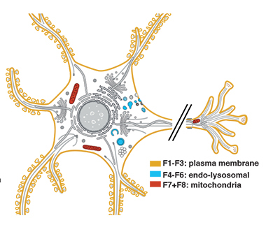
Origins, origins. Three different types of extracellular vesicle were found among different density fractions (F1-F8) from Alzheimer brain samples. Microvesicles (orange) fuse directly with the plasma membrane and are flush with cell surface proteins. Exosomes (blue) derive from endolysosomal compartments; mitovesicles (red) hail from mitochondria. [Courtesy of Fowler et al., bioRXiv, 2023.]
But do tau filaments travel this way in the human brain? To find out, co-first authors Stephanie Fowler and Tiana Behr studied tau within EVs derived from human brain samples. Using density gradient centrifugation, they fractionated EVs of various heft from frontal, temporal, and hippocampal AD brain extracts, and used mass spectrometry to probe the origins of EVs in the fractions. They identified three kinds: exosomes, which were loaded with endolysosomal proteins; microvesicles, which fuse directly with the plasma membrane and contain a glut of membrane-derived proteins; and mitovesicles, which come from mitochondria.
The scientists also detected fragments of tau, predominantly within exosomes. This tau was truncated at both the N- and C-termini and contained the third and fourth of the four microtubule binding domains. It resisted dissolution by sarkosyl and bound to antibodies for paired helical filaments, suggesting it was fibrillar.
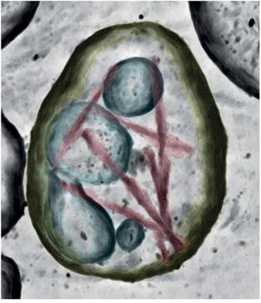
Cryo-electron tomography revealed how tau was arranged within the vesicles. Though lower resolution than cryo-EM, cryo-ET visualizes the ultrastructure and organization of proteins in situ. This is the first time the technique has been applied to human brain tissue, said Ryskeldi-Falcon. The tomographs revealed both paired helical filaments and straight filaments of tau within the lumen of EVs. Individual vesicles contained three to 50 filaments each. Compared to the micrometer-long filaments that form neurofibrillary tangles, the vesicle filaments were short, measuring 75 to 100 nanometers in length.

Bubble Tau. Tau filaments (magenta) encapsulated within extracellular vesicles touch the inner membrane (yellow) of the vesicles, and each other. They also brush up against intraluminal vesicles (blue), unknown tiny sacs seen within many EVs. [Courtesy of Fowler et al., bioRXiv, 2023.]
Strikingly, in each vesicle at least one of these stubby tau filaments was tethered to the inner membrane, while the remaining fibrils seemed to cling to the tethered filament, and/or to each other. In this way, it appears that all tau filaments may be attached—either directly or indirectly—to the inner membrane. To confirm that all the tau filaments were indeed attached in this way, the scientists extracted the membrane fraction of the vesicles. They found tau filaments still attached, while no filaments were detected among the intraluminal proteins.
Tau fibrils latched on to the membrane exclusively by their filament ends. There, flexible, elongated densities, likely representing a mysterious tethering protein, wedged in between the filament tip and the membrane (image below).
Cryo-ET also revealed smaller vesicles within the EVs. Tau filaments occasionally made contact with those (first image below). Duff does not know what these are, but said that tau filaments were never found within them, nor did tau appear tied specifically to their surface. Finally, globular entities decorated the sides of the filaments. Some of these hangers-on followed the helical symmetry of the fibril, suggesting they bound specific sequences.

Tethered Tau. Tomographs of EV cross-sections show tau filaments (pink asterisks) hitched to the luminal side of the EV membrane (yellow arrows) via unknown densities (orange arrows). [Courtesy of Fowler et al., bioRXiv, 2023.]
To Ryskeldi-Falcon, this intricate arrangement of tau filaments means that their presence is no accident. “It suggests there is something selective that’s packaging tau filaments into these vesicles,” he said.
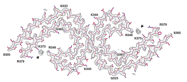
To zero in on the fibrils' atomic structure, the scientists resolved individual paired helical filaments with cryo-EM. The protofilament structure that emerged—back-to-back C-shaped protofilaments packaged with helical symmetry—was nearly identical to the one previously identified from AD brain homogenates by Michel Goedert and Sjors Scheres's groups at the MRC in Cambridge (Jul 2017 news). One notable distinction: an unknown, negatively charged molecule bridged positively charged tau residues on each tip of the C. This interloper essentially closed up the C, resulting in a more compact protofilament core (see image below).

Closing the Loop. Positioned on each end of the C-shaped protofilament near positively charged residues, negatively charged molecules (gray blob, arrow) pull the ends of the C shape closer together. [Courtesy of Fowler et al., bioRxiv, 2023.]
These EV-specific structural tweaks left tau’s ability to seed aggregates intact. The scientists found that tau filaments from EVs potently seeded tau aggregation in biosensor cell lines, and within the brains of P301S-tau transgenic mice.
Marc Diamond of UT Southwestern Medical Center in Houston called the findings “fundamentally interesting.” He cautioned that implicating EV tau in seeding or propagation should involve ruling out involvement of extravesicular tau, and testing the relative seeding efficiency of tau encased in different vesicle fractions. He thinks further context on how common these EVs with tau filaments are compared to the total population of EVs will help the field understand their biological significance.
To the authors, the findings suggest that tau filaments are actively packaged into vesicles of endolysosomal origin, which are then secreted as exosomes and, if taken up by recipient cells, may seed tauopathy. One open question is which cell types do this. Duff said this is under active investigation. She noted that neurons might be goaded into producing exosomes in response to hyperactivity brought on by exposure to Aβ oligomers.
Another suspect: viruses. The cellular response they provoke is increasingly implicated in AD, most recently with reports that viral infection ramps up EV production by neuron and that ancient viral response pathways might stoke inflammation (Apr 2023 news; Apr 2023 conference news).
Microglia may crank up EV production in response to inflammation, Duff added. Fowler said that defects in the endolysosomal system also promote the release of EVs, particularly EVs of endolysosomal origin—the type found to contain tau seeds (Cataldo et al., 2000; Hessvik et al., 2016; Abdulrahman et al., 2018).
Tsuneya Ikezu, at the Mayo Clinic in Jacksonville, Florida, was intrigued by the tethering of tau filaments to the lumen of EVs. Tau interactome studies in EVs could identify the molecules involved, and explain how the sorting works, he said. Ikezu noted that the exosomes housing the tau filaments were quite large, as exosomes go, and that tau filaments, even short ones, may be unable to squeeze inside of smaller vesicles.
Walker wondered why the fibrils happen to be conveniently sized to fit inside these vesicles. “Are they truncated segments of the much longer fibrils that typify neurofibrillary tangles, suggestive of microglial processing? Or are they growing fibrils that happen to be captured before they reach a limiting length, suggestive of neuronal origin?” Another question is whether tau filaments cross paths with oligomeric Aβ inside exosomes, since these aggregates have also been spotted within EVs (Sardar Sinha et al., 2018). “This investigation provides a model for addressing some of the many questions surrounding the pathogenicity of aberrant protein assemblies in the brain,” Walker wrote.—Jessica Shugart
References
News Citations
- Deadly Delivery: Microglia May Traffic Tau Via Exosomes
- After Eating Tangle-Tainted Neurons, Microglia Spew Tau, Lose Appetite
- From Phagocytosis to Exophagy: Microglia's Digestive Tract Dissected
- Tau Filaments from the Alzheimer’s Brain Revealed at Atomic Resolution
- Attack From Within: How Ancient Viruses Resurface to Spread Tau
- By Unleashing Microglial cGAS, Tau STINGs Neurons
Paper Citations
- Clayton K, Delpech JC, Herron S, Iwahara N, Ericsson M, Saito T, Saido TC, Ikezu S, Ikezu T. Plaque associated microglia hyper-secrete extracellular vesicles and accelerate tau propagation in a humanized APP mouse model. Mol Neurodegener. 2021 Mar 22;16(1):18. PubMed. Correction.
- Cataldo AM, Peterhoff CM, Troncoso JC, Gomez-Isla T, Hyman BT, Nixon RA. Endocytic pathway abnormalities precede amyloid beta deposition in sporadic Alzheimer's disease and Down syndrome: differential effects of APOE genotype and presenilin mutations. Am J Pathol. 2000 Jul;157(1):277-86. PubMed.
- Hessvik NP, Øverbye A, Brech A, Torgersen ML, Jakobsen IS, Sandvig K, Llorente A. PIKfyve inhibition increases exosome release and induces secretory autophagy. Cell Mol Life Sci. 2016 Dec;73(24):4717-4737. Epub 2016 Jul 20 PubMed.
- Abdulrahman BA, Abdelaziz DH, Schatzl HM. Autophagy regulates exosomal release of prions in neuronal cells. J Biol Chem. 2018 Jun 8;293(23):8956-8968. Epub 2018 Apr 26 PubMed.
- Sardar Sinha M, Ansell-Schultz A, Civitelli L, Hildesjö C, Larsson M, Lannfelt L, Ingelsson M, Hallbeck M. Alzheimer's disease pathology propagation by exosomes containing toxic amyloid-beta oligomers. Acta Neuropathol. 2018 Jul;136(1):41-56. Epub 2018 Jun 13 PubMed.
Further Reading
Papers
- Polanco JC, Li C, Durisic N, Sullivan R, Götz J. Exosomes taken up by neurons hijack the endosomal pathway to spread to interconnected neurons. Acta Neuropathol Commun. 2018 Feb 15;6(1):10. PubMed.
Annotate
To make an annotation you must Login or Register.

Comments
The University of Queensland
The work by Fowler and colleagues is beautiful, and the images visualizing tau filaments within extracellular vesicles by cryo-electron microscopy are simply stunning. I had always asked myself what would confer seeding capability to extracellular vesicles including exosomes. Is it a particular phosphorylation pattern of tau (or any other form of post-translational modification), a particular conformation or association with other proteins, or a particular aggregation state? Interestingly, this new work shows all of this.
It reveals filaments (although the discussion whether filaments or oligomers are the major toxic species is still open), there is tethering to the limiting membrane, and the enrichment with endo-lysosomal proteins hints at particular interactions and also subcellular origins. This foundational work is indeed inspiring for all of us working on tau spreading and seeding.
Emory University
Extracellular vesicles (EVs) have emerged in recent years as important vehicles of intercellular communication in many domains of biology. In the brain, EVs are produced by neurons and non-neuronal cells; their intercellular transfer can support homeostasis, but they also are capable of disseminating pathogenic protein seeds that drive neurodegenerative diseases. The once-murky details of this remarkable mechanism are now yielding to increasingly sophisticated analyses, and the characterization of polymeric tau in Alzheimer brain-derived vesicles by Fowler and colleagues is a welcome contribution.
In a series of striking ultrastructural images, they show that some vesicles contain short segments of paired helical and straight filaments of tau. These intravesicular mini-fibrils can be abundant, and their molecular structure is similar to that of the longer tau fibrils that course through the neuronal cytosol as neurofibrillary tangles. The analysis further demonstrates that fibril-containing vesicles are enriched in proteins associated with the endosomal/lysosomal system.
Which cell type(s) give rise to vesicles bearing fibrillar tau is not certain; microglia and neurons are two obvious (and not mutually exclusive) candidates. Microglia have long been known to be intimately involved in the pathobiology of AD, including Aβ plaques (e.g., Dansokho and Heneka, 2018; Walker, 2020) and tauopathy (e.g. Hansen et al., 2018; Hopp et al., 2018; Maphis et al., 2015). Previous studies have found that inhibiting microglia or the release of EVs blocks the spread of tauopathy (Asai et al., 2015).
Another potential source of the fibril-bearing vesicles is neurons themselves. In humans, the abnormal neuronal processes surrounding Aβ plaques can contain tau fibrils along with a rich variety of vesicles (“autophagic vacuoles”) that constitute an intermediate stage in lysosomal degradation (Nixon, 2007). In this regard, it is worth considering the possibility that at least some of the tau fibril-bearing vesicles derived from tissue homogenates are intracellular entities that have been released by the preparation of the samples. An electron-microscopic search for tau mini-fibrils in intracellular vesicles, especially in dystrophic neurites, might help inform this question. Leaky or ruptured dystrophic neurites still could be a source of EVs in vivo, and indeed they might account for the strong correlation between phospho-tau in the CSF and cerebral Aβ load (Therriault et al., 2023).
The researchers note that mini-fibrils appear to be end-tethered to the vesicular membrane, suggesting that they are selectively packaged, but how is it that the fibrils are conveniently sized to fit inside the vesicles? Are they truncated segments of the much longer fibrils that typify neurofibrillary tangles, suggestive of microglial processing? Or are they growing fibrils that happen to be captured before they reach a limiting length, suggestive of neuronal origin?
Finally, seeding-competent oligomeric Aβ has been found in EVs (Sardar Sinha et al., 2018). It would be interesting to know if both aberrant Aβ and tau are present in the same EVs, and if so, whether they interact in any way. This investigation provides a model for addressing some of the many questions surrounding the pathogenicity of aberrant protein assemblies in the brain.
References:
Asai H, Ikezu S, Tsunoda S, Medalla M, Luebke J, Haydar T, Wolozin B, Butovsky O, Kügler S, Ikezu T. Depletion of microglia and inhibition of exosome synthesis halt tau propagation. Nat Neurosci. 2015 Nov;18(11):1584-93. Epub 2015 Oct 5 PubMed.
Dansokho C, Heneka MT. Neuroinflammatory responses in Alzheimer's disease. J Neural Transm (Vienna). 2018 May;125(5):771-779. Epub 2017 Dec 22 PubMed.
Hansen DV, Hanson JE, Sheng M. Microglia in Alzheimer's disease. J Cell Biol. 2018 Feb 5;217(2):459-472. Epub 2017 Dec 1 PubMed.
Hopp SC, Lin Y, Oakley D, Roe AD, DeVos SL, Hanlon D, Hyman BT. The role of microglia in processing and spreading of bioactive tau seeds in Alzheimer's disease. J Neuroinflammation. 2018 Sep 18;15(1):269. PubMed.
Maphis N, Xu G, Kokiko-Cochran ON, Jiang S, Cardona A, Ransohoff RM, Lamb BT, Bhaskar K. Reactive microglia drive tau pathology and contribute to the spreading of pathological tau in the brain. Brain. 2015 Jun;138(Pt 6):1738-55. Epub 2015 Mar 31 PubMed.
Nixon RA. Autophagy, amyloidogenesis and Alzheimer disease. J Cell Sci. 2007 Dec 1;120(Pt 23):4081-91. PubMed.
Sardar Sinha M, Ansell-Schultz A, Civitelli L, Hildesjö C, Larsson M, Lannfelt L, Ingelsson M, Hallbeck M. Alzheimer's disease pathology propagation by exosomes containing toxic amyloid-beta oligomers. Acta Neuropathol. 2018 Jul;136(1):41-56. Epub 2018 Jun 13 PubMed.
Therriault J, Vermeiren M, Servaes S, Tissot C, Ashton NJ, Benedet AL, Karikari TK, Lantero-Rodriguez J, Brum WS, Lussier FZ, Bezgin G, Stevenson J, Rahmouni N, Kunach P, Wang YT, Fernandez-Arias J, Socualaya KQ, Macedo AC, Ferrari-Souza JP, Ferreira PC, Bellaver B, Leffa DT, Zimmer ER, Vitali P, Soucy JP, Triana-Baltzer G, Kolb HC, Pascoal TA, Saha-Chaudhuri P, Gauthier S, Zetterberg H, Blennow K, Rosa-Neto P. Association of Phosphorylated Tau Biomarkers With Amyloid Positron Emission Tomography vs Tau Positron Emission Tomography. JAMA Neurol. 2023 Feb 1;80(2):188-199. PubMed.
Walker LC. Aβ Plaques. Free Neuropathol. 2020;1 Epub 2020 Oct 30 PubMed.
Mayo Clinic Florida
This study demonstrates for the first time the presence of PHF or SF of tau, which are the truncated form encompassing residues 305-379 and detected as 12 or 24 kD by TauC. The authors beautifully show the structure of PHF by cryo-electron tomography and single-particle cryo-EM, which also revealed the structure of PHF as two C-shaped protofilaments and interesting anionic molecules between residue R349 and K375.
The study also demonstrates that AD brain-derived EVs have tau seeding activity both in vitro and in vivo. This is a very interesting finding and provides evidence that brain-derived EVs carry PHF tau seeds capable of tau propagation.
We detected tau protein by Tau13 antibody, which may be attributed to the difference in the isolation method of EVs, sensitivity of the WB technique, or loading volume of the EV samples. We also detected globular tau aggregates in brain EVs (Ruan et al., 2021), which may also be present in Fowler et al.'s preparation.
The authors also show tethering of PHF to the lumen of EVs at the tail end of PHF, suggesting the presence of a unique tau-interacting molecule for the insertion of PHF tau to the EVs. Tau interactome studies will be useful for the identification of the tau interaction molecule for the molecular understanding of the sorting system.
References:
Ruan Z, Pathak D, Venkatesan Kalavai S, Yoshii-Kitahara A, Muraoka S, Bhatt N, Takamatsu-Yukawa K, Hu J, Wang Y, Hersh S, Ericsson M, Gorantla S, Gendelman HE, Kayed R, Ikezu S, Luebke JI, Ikezu T. Alzheimer's disease brain-derived extracellular vesicles spread tau pathology in interneurons. Brain. 2021 Feb 12;144(1):288-309. PubMed. Correction.
UCLA
Scientists have long postulated that pathological tau fibrils spread throughout the brain in a prion-like fashion using intercellular transfer mediated by extracellular vesicles (EVs). Benjamin Falcon’s lab in the MRC Laboratory of Molecular Biology now reports isolation of EVs from AD patients and shows intercellular tau transport in action.
In this preprint, the authors isolated EV fractions from postmortem AD patient brain tissues that contain microvesicles, exosomes, and the newly discovered mitovesicles. After confirming the presence of sarkosyl insoluble tau in the EVs, the authors performed electron cryotomography (cryo-ET) on these freshly extracted vesicles. Cryo-ET combines images of the specimen as it is rotated in the electron beam, producing a picture of the tau fibers inside intact vesicles. Averaging images (“subtomogram averaging”) of fibers inside EVs gave low-resolution densities resembling tau paired helical filaments (PHFs) and straight filaments (SFs).
Subsequent single-particle cryo-EM performed on extracted fibers confirmed the identity of tau PHFs inside EVs. The only difference between the EV-extracted tau PHFs and other previously solved PHF structures from tau extracted from total brain homogenates is the presence of an additional density between the positively charged side chains of Arginine 349 and Lysine 375, giving this PHF an ever-so-slightly-more-compact C-shaped fold.
While studying the tomograms of tau containing EVs, the authors detected an elongated density tethering tau filament ends to the surrounding membrane. It would appear that not all filaments are tethered. Rather, most filaments make lateral contacts with the tethered filament and each other, resulting in bundles of tau filaments enveloped by the EV. Tau was found in the membrane fraction following EV fractionation, further demonstrating direct membrane interaction between tau and the EV membrane.
Whereas these findings may suggest hypotheses about tau being actively sorted into vesicles, and may point to new therapeutic targets, the identity and contact points of the postulated tethers are not yet established. Overall, this study elevates ex vivo tau studies by placing tau directly in a cellular context. It also shows the promise of in situ cryo-ET to study neurodegeneration in a native biological environment.
—Xinyi Cheng is co-author of this comment.
Make a Comment
To make a comment you must login or register.