Sans Plexin-B1, Glial Net Tightens Around Plaques
Quick Links
Sometimes being polite does more harm than good. When glia surround amyloid plaques, they keep their distance from one another in structured cellular nets. These manners are enforced by the axon guidance receptor Plexin-B1, according to a paper published May 27 in Nature Neuroscience. Scientists led by Bin Zhang, Hongyan Zou, and Roland Friedel at New York’s Icahn School of Medicine at Mount Sinai reported that deleting Plexin-B1 in a mouse model of amyloidosis freed astrocytes and microglia to huddle closer around plaques. This led to smaller, denser aggregates that posed less of a threat to nearby synapses. Unfettering the cells of their social graces also dampened neuroinflammation and lessened memory loss in the mice.
- Plexin-B1 expression by astrocytes imposes cell spacing in glial nets.
- Without Plexin-B1, glia huddle more closely together, and with plaques.
- The result? Smaller plaques, protected synapses, spared memory. In mice.
The authors, and other scientists, had previously tied elevated Plexin-B1 expression in postmortem brain samples to cognitive decline and AD (Wang et al., 2018; Jun 2018 news). However, how this association might work remained obscure. Plexin-B1 binds semaphorin ligands, including Sema4D. They sit on the surface of multiple neural cell types, but can also be secreted. These receptor-ligand pairs guide axon growth during development, and have also been found to repel cells from one another, presumably giving them enough “personal space” to get on with their jobs (Roy et al., 2017; Zhou et al., 2020).

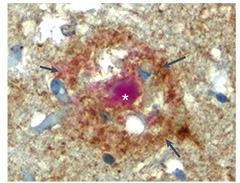
Plexin Corona. Amyloid plaques (crimson) surrounded by Plexin-B1 (brown) in a postmortem brain sample from a person with AD. [Courtesy of Huang et al., Nature Neuroscience, 2024.]
To investigate if Plexin-B1 influences AD pathogenesis, co-first authors Yong Huang and Minghui Wang first asked which cell types make the receptor. Their previous work suggested astrocytes were the main source, and a deeper analysis of data from Mount Sinai’s Brain Bank AD cohort showed that indeed, expression of Plexin-B1 in astrocytes, but not other cell types, correlated with plaque burden. Immunohistochemistry of AD brain tissue revealed plaques encircled by a prominent corona of Plexin B1, consistent with the shape of a glial net (image at right), while expression was weak far from plaques. Notably, among samples from 11 people who spanned the spectrum of AD, Plexin-B1 expression surrounding plaques was highest among those with the heaviest overall burden of AD pathology based on amyloid plaques, neuritic plaques, and neurofibrillary tangles. In APP/PS1 mice, plaque-adjacent astrocytes, but not microglia, expressed the receptor.
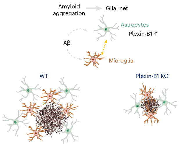
To find out if the Plexin-B1 around plaques plays any role in pathology, the researchers knocked out the receptor from APP/PS1 mice. The results were striking. In 6-month-old APP/PS1 control mice, astrocytes formed spheres around plaques that were about two cell layers wide. In mice lacking Plexin-B1, the footprint of this net cinched by more than half. It contained fewer astrocytes, but closer together, and more of them directly probed the plaque surface with their processes. Microglia responded similarly, moving closer to each other and to plaques, though again with fewer cells recruited overall.

Tightening Net. In APP/PS1 mice (left), astrocytes (green), and microglia (light grey) encircle an Aβ plaque (red) in a loose net. In Plexin-B1 knockouts (right), the net tightens, and glia directly engage the plaque surface. [Courtesy of Huang et al., Nature Neuroscience, 2024.]
Why the packed net? Single-cell RNA sequencing revealed nine subclusters of astrocytes, including one with a reactive, disease-associated astrocyte (DAA) signature. In APP/PS1 controls, these cells expressed most of the Plexin-B1, and the scientists think these are the astrocytes surrounding plaques. Without Plexin-B1, this cluster shrank, and the cells expressed higher levels of genes involved in forming cell projections and bolstering energy metabolism. Plexin-B1 deletion also reduced numbers of disease-associated microglia (DAM), while boosting their expression of genes involved in microglial activation, chemoattraction, host defense, and antigen presentation. In all, the findings hinted at a scaled-down, yet more potent, response to plaques among microglia and astrocytes. Notably, communication between the two cell types seemed to improve, as expression of both ligands and receptors involved in cell-to-cell signaling surged.

Huddle Up! In APP/PS1 controls (left), astrocytes (green) and microglia (brown) form a wide glial net around plaques. Without Plexin-B1 to enforce cell spacing (right), glia crowd together and around plaques, compacting them more. [Courtesy of Huang et al., Nature Neuroscience, 2024.]
This potency manifested in the plaque load. Without Plexin-B1, average plaque size shrank by almost half. They also became denser. In APP/PS1 controls, half of the plaques were diffuse, 15 percent had dense cores, and the rest were of mixed composition. The opposite was true in Plexin-B1 knockouts, where half of the plaques had dense cores, and 15 percent were purely diffuse. This greater compaction spared nearby synapses, as seen by a 50 percent reduction in dystrophic neurites. Finally, this translated into better working memory for the mice, which more readily learned the location of an escape hatch on a circular platform relative to their Plexin-B1-replete counterparts.
The shift toward more dense-core plaques jibes with previous studies implicating microglia as plaque compactors (Apr 2021 news; May 2016 news). With microglia better able to do this, fewer glia need to be recruited to the scene, leading to fewer cells surrounding plaques and less neuroinflammation.
Zhang told Alzforum that details about the signaling pathway need to be worked out. Greg Lemke of the Salk Institute in La Jolla, California, agrees, but found the study interesting. “Important features of signaling within the glial nets—e.g., the identity of the Plexin-B1 ligand, how Plexin-B1 activation in astrocytes affects the physiology of microglia—remain to be determined,” he wrote. “Nonetheless, pharmacological perturbation of Plexin-B1 signaling could be experimentally assessed as an AD therapy.”
A recent study suggests that microglia expressing Sema4D can activate Plexin-B1 on astrocytes (Clark et al., 2021). Indeed, some Plexin-B1 ligands—the semaphorins—are already being targeted in clinical trials. For example, a Sema4D-targeted antibody, pepinemab, is being tested in clinical trials for AD, based in part on observations that neurons upregulate Sema4D in AD (Evans et al., 2022). A Sema4D-blocking nanobody, as well as a Plexin-B1-binding peptide, has also been developed (Cowan et al., 2023; Matsunaga et al., 2016).—Jessica Shugart
References
News Citations
- Culling Connection From Chaos, Alzheimer’s Genetic Network Study Pins PLXNB1 and INPPL1
- Microglia Build Plaques to Protect the Brain
- Barrier Function: TREM2 Helps Microglia to Compact Amyloid Plaques
Therapeutics Citations
Paper Citations
- Wang M, Beckmann ND, Roussos P, Wang E, Zhou X, Wang Q, Ming C, Neff R, Ma W, Fullard JF, Hauberg ME, Bendl J, Peters MA, Logsdon B, Wang P, Mahajan M, Mangravite LM, Dammer EB, Duong DM, Lah JJ, Seyfried NT, Levey AI, Buxbaum JD, Ehrlich M, Gandy S, Katsel P, Haroutunian V, Schadt E, Zhang B. The Mount Sinai cohort of large-scale genomic, transcriptomic and proteomic data in Alzheimer's disease. Sci Data. 2018 Sep 11;5:180185. PubMed.
- Deb Roy A, Yin T, Choudhary S, Rodionov V, Pilbeam CC, Wu YI. Optogenetic activation of Plexin-B1 reveals contact repulsion between osteoclasts and osteoblasts. Nat Commun. 2017 Jun 21;8:15831. PubMed.
- Zhou X, Wahane S, Friedl MS, Kluge M, Friedel CC, Avrampou K, Zachariou V, Guo L, Zhang B, He X, Friedel RH, Zou H. Microglia and macrophages promote corralling, wound compaction and recovery after spinal cord injury via Plexin-B2. Nat Neurosci. 2020 Mar;23(3):337-350. PubMed.
- Clark IC, Gutiérrez-Vázquez C, Wheeler MA, Li Z, Rothhammer V, Linnerbauer M, Sanmarco LM, Guo L, Blain M, Zandee SE, Chao CC, Batterman KV, Schwabenland M, Lotfy P, Tejeda-Velarde A, Hewson P, Manganeli Polonio C, Shultis MW, Salem Y, Tjon EC, Fonseca-Castro PH, Borucki DM, Alves de Lima K, Plasencia A, Abate AR, Rosene DL, Hodgetts KJ, Prinz M, Antel JP, Prat A, Quintana FJ. Barcoded viral tracing of single-cell interactions in central nervous system inflammation. Science. 2021 Apr 23;372(6540) PubMed.
- Evans EE, Mishra V, Mallow C, Gersz EM, Balch L, Howell A, Reilly C, Smith ES, Fisher TL, Zauderer M. Semaphorin 4D is upregulated in neurons of diseased brains and triggers astrocyte reactivity. J Neuroinflammation. 2022 Aug 6;19(1):200. PubMed.
- Cowan R, Trokter M, Oleksy A, Fedorova M, Sawmynaden K, Worzfeld T, Offermanns S, Matthews D, Carr MD, Hall G. Nanobody inhibitors of Plexin-B1 identify allostery in plexin-semaphorin interactions and signaling. J Biol Chem. 2023 Jun;299(6):104740. Epub 2023 Apr 23 PubMed.
- Matsunaga Y, Bashiruddin NK, Kitago Y, Takagi J, Suga H. Allosteric Inhibition of a Semaphorin 4D Receptor Plexin B1 by a High-Affinity Macrocyclic Peptide. Cell Chem Biol. 2016 Nov 17;23(11):1341-1350. Epub 2016 Oct 27 PubMed.
Further Reading
No Available Further Reading
Primary Papers
- Huang Y, Wang M, Ni H, Zhang J, Li A, Hu B, Junqueira Alves C, Wahane S, Rios de Anda M, Ho L, Li Y, Kang S, Neff R, Kostic A, Buxbaum JD, Crary JF, Brennand KJ, Zhang B, Zou H, Friedel RH. Regulation of cell distancing in peri-plaque glial nets by Plexin-B1 affects glial activation and amyloid compaction in Alzheimer's disease. Nat Neurosci. 2024 Aug;27(8):1489-1504. Epub 2024 May 27 PubMed.
Annotate
To make an annotation you must Login or Register.

Comments
Salk Institute
This interesting study provides evidence that the class 4 semaphorin receptor Plexin-B1, which is prominently expressed by the astrocytes that surround amyloid beta plaques in Alzheimer’s disease, regulates both astrocyte spacing around, and microglia association with, these plaques.
When germline Plexin-B1 mouse mutants are crossed into the APP/PS1 AD model, microglia and peripheral astrocytes are seen to associate much more tightly with Aβ plaques, and the cytoarchitecture of these plaques is seen to shift from a wispy fibrillar (more neurotoxic) to a compacted dense-core (less neurotoxic) configuration.
Important features of signaling within the glial nets—e.g., the identity of the Plexin-B1 ligand, how Plexin-B1 activation in astrocytes affects the physiology of microglia—remain to be determined. Nonetheless, pharmacological perturbation of Plexin-B1 signaling—not addressed in this study—could be experimentally assessed as an AD therapy.
Vaccinex, Inc.
As noted in this interesting article, we are testing a Sema4D-targeted antibody, pepinemab, in an early stage randomized, Phase 1b/2 clinical trial for AD. SEMA4D is a major ligand for activation of plexin-B1; it is, therefore, of considerable interest to compare the effects of anti-SEMA4D antibody blockade with those reported by the present authors for plexin-B1 KO.
The effect of plexin-B1 KO on the glial net surrounding amyloid plaques, including interactions among astrocytes and between astrocytes and microglia, is certainly interesting and, as suggested by the authors, may contribute to relieving AD pathology. An important property of astrocytes is their distribution in tiled fashion such that individual astrocytes establish a local sphere of influence in which surveillance is enabled through numerous branching projections. Remarkably, a single human astrocyte may in this fashion make hundreds of thousands of cell contacts. This contrasts with the mobile surveillance strategy adopted by less numerous microglia and implies a mechanism for mutual repulsion by astrocytes. Results of the present report suggest that plexin-B1 and an unidentified ligand, possibly SEMA4D, may serve this purpose. The same, or similar, ligand-receptor signaling pairs may also mediate crosstalk between astrocytes and microglia as previously suggested by Clark et al., 2021, who identified SEMA4D as a relevant ligand.
It is, however, important to note that these are not the only astrocyte interactions mediated by these, and potentially related, ligand-receptor signaling pairs, e.g. plexin-B2 and other class IV semaphorins. We have reported that SEMA4D is highly upregulated in neurons during progression of AD and Huntington’s disease (HD), and that this facilitates signaling between neurons and closely associated plexin-B1 positive astrocytes which, in their homeostatic state, are responsible for recycling glutamate at synapses, support energy metabolism, and maintain ionic gradients (Evans et al., 2022). This SEMA4D-plexin-B1 interaction appears to trigger reactive astrogliosis, including loss of supportive functions and gain of neuroinflammatory activity. We have reported beneficial effects of treatment with SEMA4D blocking antibody in HD (Feigin et al., 2022) and plan to shortly report topline data for the SIGNAL-AD randomized, Phase 1b/2 study in AD.
Unfortunately, for what appear to be technical reasons, neither Huang et al., 2024, nor Clark et al., 2021, were able to characterize astrocyte interactions with neurons. Zhang at al. comment that “attempts for additional detection of Plexin-B1 in human brain specimens by RNAscope ISH or IF were not successful, likely due to preservation challenges for postmortem tissues,” and "due to technical factors related to tissue dissociation, neurons were underrepresented in this analysis.” As noted, peri-plaque astrocytes are a minority population compared to the general astrocyte populations in plaque-free areas. We wish to highlight that these other, more numerous, astrocyte populations can contribute to neuroinflammation and neurodegeneration in different ways. Indeed, the interesting thing about targeting central regulators of glial physiology, like plexin-B1 and SEMA4D, is that their effects are pleiotropic.
References:
Huang Y, Wang M, Ni H, Zhang J, Li A, Hu B, Junqueira Alves C, Wahane S, Rios de Anda M, Ho L, Li Y, Kang S, Neff R, Kostic A, Buxbaum JD, Crary JF, Brennand KJ, Zhang B, Zou H, Friedel RH. Regulation of cell distancing in peri-plaque glial nets by Plexin-B1 affects glial activation and amyloid compaction in Alzheimer's disease. Nat Neurosci. 2024 Aug;27(8):1489-1504. Epub 2024 May 27 PubMed.
Clark IC, Gutiérrez-Vázquez C, Wheeler MA, Li Z, Rothhammer V, Linnerbauer M, Sanmarco LM, Guo L, Blain M, Zandee SE, Chao CC, Batterman KV, Schwabenland M, Lotfy P, Tejeda-Velarde A, Hewson P, Manganeli Polonio C, Shultis MW, Salem Y, Tjon EC, Fonseca-Castro PH, Borucki DM, Alves de Lima K, Plasencia A, Abate AR, Rosene DL, Hodgetts KJ, Prinz M, Antel JP, Prat A, Quintana FJ. Barcoded viral tracing of single-cell interactions in central nervous system inflammation. Science. 2021 Apr 23;372(6540) PubMed.
Evans EE, Mishra V, Mallow C, Gersz EM, Balch L, Howell A, Reilly C, Smith ES, Fisher TL, Zauderer M. Semaphorin 4D is upregulated in neurons of diseased brains and triggers astrocyte reactivity. J Neuroinflammation. 2022 Aug 6;19(1):200. PubMed.
Feigin A, Evans EE, Fisher TL, Leonard JE, Smith ES, Reader A, Mishra V, Manber R, Walters KA, Kowarski L, Oakes D, Siemers E, Kieburtz KD, Zauderer M, Huntington Study Group SIGNAL investigators. Pepinemab antibody blockade of SEMA4D in early Huntington's disease: a randomized, placebo-controlled, phase 2 trial. Nat Med. 2022 Oct;28(10):2183-2193. Epub 2022 Aug 8 PubMed. Correction.
Make a Comment
To make a comment you must login or register.