Receptor Decoy Raises Risk of Alzheimer’s—But Only in Women
Quick Links
Women are at higher risk than men of developing AD, and their dementia advances more quickly after diagnosis. Scientists led by Nancy Ip, Hong Kong University of Science and Technology, blame soluble ST2, a protein that dampens microglial activation and phagocytosis, for some of this difference.
- A soluble form of the IL-33 receptor, sST2, blocks activation of microglia.
- A genetic variant that suppresses sST2 associates with reduced plaque load.
- Mice injected with sST2 poorly phagocytosed plaques.
In the July 15 Nature Aging, they reported that female APOE4 carriers who had high levels of sST2 in their blood and cerebrospinal fluid were likelier to have AD. They had more amyloid plaques and more shrunken brains. A Mendelian randomization analysis suggested that a variant that suppresses sST2 expression slows brain atrophy and cognitive decline, and reduces AD risk. These findings may explain some of the heterogeneity of AD risk between the sexes, and implicate a new pathway in microglial dysfunction.
“If these results are validated in additional well-characterized, longitudinal cohort studies, sST2 might be an interesting new drug target, especially for women with AD,” Oskar Hansson, Lund University, Sweden, wrote to Alzforum (full comment below).

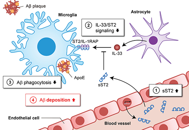
Decoy Receptor. In APOE4 carriers, brain epithelial cells produce sST2 (1, blue squiggle), which binds to IL-33 before the interleukin can bind ST2L (2, blue/purple pair), its receptor on microglia. The sST2 decoy suppresses phagocytosis of Aβ (3), increasing plaque load (4). [Courtesy of Nancy Ip.]
Soluble ST2 comprises the extracellular domain of ST2L, a cell-surface IL-33 receptor on microglia. Unlike other extracellular domains such as soluble TREM2 and soluble APP, sST2 is not shed by a protease. Instead, the protein is transcribed from IL1RL1, the gene that encodes ST2L. In essence, sST2 and ST2L are transcribed separately from the same gene. As a result, sST2 can act as a decoy, sequestering IL-33 and preventing it from binding its microglial receptor (reviewed by Pusceddu et al., 2019).
This could be important for AD and related diseases, since IL-33/ST2L signaling alerts microglia to tissue damage, prompting them to clean up plaques and prune synapses (Lau et al., 2020; Jul 2020 news; Feb 2018 news). Ip and others have reported high levels of sST2 in the blood of people with mild cognitive impairment or early-stage AD (Fu et al., 2016; Saresella et al., 2020).
To investigate this relationship, first author Yuanbing Jiang correlated plasma and cerebrospinal fluid sST2 levels with markers of AD pathology in three cohorts: 345 Chinese people with AD and 345 controls; 107 people with AD from the Stanford Alzheimer’s Disease Research Center; and 75 AD cases in the U.K. Brain Banks Network (UKBBN). Compared to controls, people with AD had higher plasma and CSF sST2, which correlated with greater cortical amyloid load, worse gray-matter shrinkage, and higher plasma phosphotau-181 and neurofilament light.
These associations were strongest in women, especially APOE4 carriers. Women also had a steeper increase in sST2 with age than did men.
Variants in IL1RL1 have been reported to shift plasma sST2 levels (Ho et al., 2013). Could such mutations explain some of the difference in sST2 levels? Whole-genome sequencing of the Chinese cohort found 575 variants in the genome that associated with plasma sST2, 79 of which were in or near IL1RL1. One—the noncoding mutation rs1921622—most strongly correlated with sST2. Homozygous carriers had 50 percent less sST2 in plasma and 75 percent less in CSF.
The data suggested that the variant reduces sST2 expression. The scientists checked this by analyzing the GTEx dataset and a single-nucleus RNA-sequencing dataset of frontal cortex AD tissue that they had previously obtained (see Oct 2020 news). Carriers of rs1921622 expressed less sST2, but not ST2L, than did noncarriers. Notably, the snRNA-Seq data revealed that only brain endothelial cells expressed sST2. This suggests a new pathogenic role of the brain vasculature in AD, the authors wrote.
Does this variant reduce AD risk? Using Mendelian randomization, a statistical tool that distinguishes causative from correlative relationships between a trait and a disease, the scientists analyzed genetic data from 5,910 people with AD and 5,477 controls from three Chinese datasets and six European ancestry-based cohorts, combined. Indeed, compared to female APOE4 carriers without the rs1921622 variant, those with the allele had less sST2 and a 25 percent lower chance of having AD. The variant also associated with larger entorhinal cortices, less gray-matter atrophy, better cognitive test scores, and delayed dementia onset.
What about brain amyloid? Immunohistochemistry of frontal cortex tissue from 42 women and 36 men with AD from the UKBBN showed that female, but not male, rs1921622 carriers accumulated fewer plaques and had more plaque-associated microglia than noncarriers, but only if they also carried an APOE4 allele (see image below).
Ip’s snRNA-seq dataset of AD frontal cortex showed that female APOE4 carriers with the variant expressed more microglial genes involved in Aβ clearance, including APOE, and downregulated more homeostatic genes than did noncarriers, suggesting that the variant supports microglial activation.

Protective Variant. Women who had had AD and carried an APOE4 allele had more plaques (brown) and fewer plaque-associated microglia (purple) in the frontal cortex (left) than did APOE4 carriers who had the sST2 variant, rs1921622 (right). [Courtesy of Jiang et al., Nature Aging, 2022.]
To test sST2-microglial interactions in vivo, the scientists infused the protein into the cerebral ventricles of 3-month-old 5xFAD mice for 28 days. Females deposited more filamentous amyloid plaques in their cortices, though the total number of plaques was unchanged. Ip thinks that sST2 keeps microglia from surrounding and compacting plaques into a less toxic form, as has been reported (Jun 2020 news; Jan 2019 news; May 2016 news). Indeed, the microglia exposed to sST2 covered less plaque surface area, and fewer of them contained amyloid. To the authors, this implies that sST2 does not prevent microglial migration to plaques but hampers their phagocytosis.
Renzo Mancuso, University of Antwerp, Belgium, agreed. “At first glance, infusion of sST2 seems to boost microglial activity with more microglia around plaques, but a closer look shows that they may not be able to interact with amyloid fibrils adequately,” he wrote (full comment below).
All told, women APOE4 carriers whose blood and CSF sST2 levels are high have impaired microglia and higher odds of AD, while those with the rs1921622 variant are somewhat protected.
Why the effects of IL-33/ST2L signaling are linked to sex and APOE genotype is unclear. Perhaps testosterone, which ramps up IL-33/ST2 signaling, protects men from the deleterious effects of sST2? As for APOE4, the authors think that IL-33/ST2 signaling may control microglial activation differently in people with different APOE genotypes.—Chelsea Weidman Burke
References
News Citations
- With IL-33, Neurons Tempt Microglia to Nibble At Synapses
- Astrocytic IL-33 Signals Microglia to Engulf Synapses
- Do Endothelial Cells Spur Capillaries to Grow in Alzheimer’s Brain?
- In Mice, Activating TREM2 Tempers Plaque Toxicity, not Load
- Without TREM2, Plaques Grow Fast in Mice, Have Less ApoE
- Barrier Function: TREM2 Helps Microglia to Compact Amyloid Plaques
Research Models Citations
Paper Citations
- Pusceddu I, Dieplinger B, Mueller T. ST2 and the ST2/IL-33 signalling pathway-biochemistry and pathophysiology in animal models and humans. Clin Chim Acta. 2019 Aug;495:493-500. Epub 2019 May 25 PubMed.
- Lau SF, Chen C, Fu WY, Qu JY, Cheung TH, Fu AK, Ip NY. IL-33-PU.1 Transcriptome Reprogramming Drives Functional State Transition and Clearance Activity of Microglia in Alzheimer's Disease. Cell Rep. 2020 Apr 21;31(3):107530. PubMed.
- Fu AK, Hung KW, Yuen MY, Zhou X, Mak DS, Chan IC, Cheung TH, Zhang B, Fu WY, Liew FY, Ip NY. IL-33 ameliorates Alzheimer's disease-like pathology and cognitive decline. Proc Natl Acad Sci U S A. 2016 May 10;113(19):E2705-13. Epub 2016 Apr 18 PubMed.
- Saresella M, Marventano I, Piancone F, La Rosa F, Galimberti D, Fenoglio C, Scarpini E, Clerici M. IL-33 and its decoy sST2 in patients with Alzheimer's disease and mild cognitive impairment. J Neuroinflammation. 2020 Jun 6;17(1):174. PubMed.
- Ho JE, Chen WY, Chen MH, Larson MG, McCabe EL, Cheng S, Ghorbani A, Coglianese E, Emilsson V, Johnson AD, Walter S, Franceschini N, O'Donnell CJ, CARDIoGRAM Consortium, CHARGE Inflammation Working Group, Dehghan A, Lu C, Levy D, Newton-Cheh C, CHARGE Heart Failure Working Group, Lin H, Felix JF, Schreiter ER, Vasan RS, Januzzi JL, Lee RT, Wang TJ. Common genetic variation at the IL1RL1 locus regulates IL-33/ST2 signaling. J Clin Invest. 2013 Oct;123(10):4208-18. Epub 2013 Sep 3 PubMed.
External Citations
Further Reading
Primary Papers
- Jiang Y, Zhou X, Wong HY, Ouyang L, FCF Ip, Chau VMN, Lau SF, Wu W, Wong DYK, Seo H, Fu WY, Lai NCH, Chen Y, Chen Y, Tong EPS, Alzheimer’s Disease Neuroimaging Initiative, Mok VCT, Kwok TCY, Mok KY, Shoai M, Lehallier B, Losada PM, O’Brien E, Porter T, Laws SM, Hardy J, Wyss-Coray T, Masters CL, Fu AKY, Ip NY. An IL1RL1 genetic variant lowers soluble ST2 levels and the risk effects of APOE-ε4 in female patients with Alzheimer’s disease. Nat Aging (2022). Nature Aging
Annotate
To make an annotation you must Login or Register.

Comments
VIB-Center for Molecular Neurology
This comprehensive paper goes from clinical observations and genetics to proof of concept in experimental models. One exciting thing about it: It showcases how complicated Alzheimer’s disease is. The authors describe cross talk between endothelial cells and microglia, which has the potential to modulate microglial activity and Aβ deposition.
They also describe genetic variants that can alter this particular interaction and, more importantly, the penetrance of APOE4/4 in AD subjects. This is important in light of available data from my recent preprint and the recent Cell paper from the Goate lab showing that APOE4/4 alone in microglia is perhaps not the most determining factor, implying that there must be other genetic/cellular interactions that play a crucial role.
The cherry on top of the cake is that the effect depends on gender. According to the experimental data shown here, this seems to be dictated by gender differences in the microglia, as the injection of sST2 results in diminished plaque deposition only in female mice.
Regarding the microglial part of the study, I think it is a proof of concept showcasing the multicellular nature of the immune response that occurs in the Alzheimer’s brain. At first glance, infusion of sST2 seems to boost microglia activity with more microglia around plaques, but a closer look shows that they may be unable to adequately interact with amyloid fibrils. It would be interesting to dig deeper into what is the phenotype and surface expressome of these cells, as there we may find key molecular players that mediate the interaction and clearance of amyloid deposits.
Lund University
This study is comprehensive and impressive. If the main results of the study are validated in additional well-characterized, longitudinal cohort studies, sST2 might be an interesting new drug target, especially for females with AD. To me, it would be very interesting to study whether sST2 levels in CSF or plasma are associated with accumulation of Aβ or tau aggregates over time in females with preclinical or prodromal AD.
National Institute on Aging
National Institute on Aging
These findings highlight an important endogenous mechanism capable of influencing Aβ clearance among those most at risk of dementia, namely female APOEε4 carriers. Through an integrative and comprehensive multidisciplinary approach, this work led by Nancy Ip’s group shows how lower circulating levels of the sST2 decoy receptor (due to a SNP in its IL1RL1 gene) can promote a microglia activation state, promote microglial clearance of Aβ from the brain, and reduce AD risk among women harboring an APOEε4 allele.
As we noted in our commentary (Duggan and Walker, 2022) published alongside this article in Nature Aging, there is much to be learned by understanding what factors explain the sex- and APOE genotype-specific associations observed by the authors. One possibility is that CSF sST2 levels only influence microglial responses to brain Aβ in individuals who have the combination of high cortical Aβ and suboptimal microglia function—two features more likely to occur in female APOEε4 carriers. Understanding the relationship between sST2 levels and microglia functioning across various disease stages may be informative in this regard and could shed additional light on the potential utility of sST2 as a drug target.
Another outstanding question concerns the mechanistic underpinnings of this receptor’s effects. Does this secreted decoy receptor indeed trigger microglial clearance of Aβ via the canonical signaling cascade for which it is a decoy (i.e., IL-33-ST2L)? In other words, are the low levels of sST2 and associated decreases in Aβ attributed to increased IL-33-ST2L signaling in microglia, which is otherwise inhibited by high levels of sST2? Or, like some other decoy receptors, might sST2 influence microglial clearance of Aβ by other mechanisms independent of its canonical signaling pathway, such as binding directly to Aβ and regulating its uptake (Chakrabarty et al., 2018; Liu et al., 2017)?
Illuminating such mechanistic bases of sST2 may enable the development of therapeutics that boost its protective effects. For example, if the absence of this secreted decoy receptor indeed triggers microglial Aβ clearance by facilitating its canonical signaling cascade (i.e., IL-33-ST2L), then allosteric modulators which enhance IL-33-ST2L binding may augment the beneficial effects associated with lower sST2 levels. Ultimately, Jiang et al.'s findings draw attention to a potentially novel circulating biomarker as well as a molecular target for AD that calls for future investigation.
References:
Duggan MR, Walker KA. Reducing decoys focuses fighting microglia. Nat Aging, July 2022 Nature Aging
Chakrabarty P, Li A, Ladd TB, Strickland MR, Koller EJ, Burgess JD, Funk CC, Cruz PE, Allen M, Yaroshenko M, Wang X, Younkin C, Reddy J, Lohrer B, Mehrke L, Moore BD, Liu X, Ceballos-Diaz C, Rosario AM, Medway C, Janus C, Li HD, Dickson DW, Giasson BI, Price ND, Younkin SG, Ertekin-Taner N, Golde TE. TLR5 decoy receptor as a novel anti-amyloid therapeutic for Alzheimer's disease. J Exp Med. 2018 Sep 3;215(9):2247-2264. PubMed.
Liu YL, Chen WT, Lin YY, Lu PH, Hsieh SL, Cheng IH. Amelioration of amyloid-β-induced deficits by DcR3 in an Alzheimer's disease model. Mol Neurodegener. 2017 Apr 24;12(1):30. PubMed.
Make a Comment
To make a comment you must login or register.