Mesh, not Membrane: Pia Filters CSF, Gets Clogged by Aβ Fibrils
Quick Links
Scientists have long believed that the pia, the innermost of the three meningeal membranes that surround the brain, is impermeable to cerebrospinal fluid. But the pia has been hard to study. This delicate tissue cannot easily be imaged, and there are no pia-specific antibodies. Now, researchers led by Rupal Mehta, Rush University, Chicago, challenge the long-held view. Using high-resolution imaging and a new marker, they found that the pia is more like a net than a membrane, at least in mice. In the July 6 Nature Communications, they report that the mesh-like network encases brain-penetrating blood vessels, creating perivascular spaces that filter CSF and act as scaffolds for macrophages that surveil solutes entering the brain.
- In mice, the perivascular pia houses macrophages, perhaps helping them surveil the CSF.
- It thins as mice age, allowing CSF surrounding the meninges to seep into brain tissue.
- In amyloidosis mice, the pia thickens, trapping Aβ fibrils and curtailing CSF flow.
As the mice aged, their delicate pia disintegrated, allowing CSF from the meninges to flow into the parenchyma. In contrast, mouse models of amyloidosis developed a thick pia network as they aged, which trapped Aβ and curtailed CSF flow. “Our work fundamentally alters the understanding of meningeal and cerebrovascular anatomy,” the authors wrote.
“This study shows greater complexity of pia and vascular anatomy than previously thought, revealing that the perivascular space does not prevent the trafficking of molecules through the CSF,” Costantino Iadecola at Weill Cornell Medical College, New York, told Alzforum. Richard Daneman, University of California, San Diego, agreed. “These findings will help researchers better understand fluid dynamics in the brain and how they change with aging and in Alzheimer's disease,” he said.

Porous Pia. Processes (green) of pial cells (blue nuclei) intermingle to form a porous network, rather than a solid membrane. [Courtesy of Mestre et al., Nature Communications, 2022.]
To get a closer look at the pia, first author Humberto Mestre, now at the University of Rochester, New York, labeled slices from the cerebral cortices of 2-month-old, wild-type mice with a fluorescent antibody to ERTR7, a recently recognized pia cell marker (Hannocks et al., 2018).
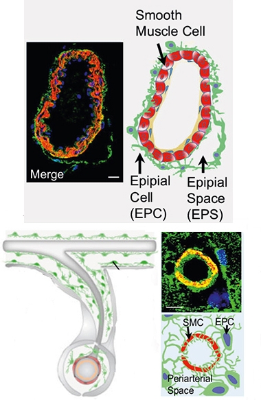
Under ultrastructural light microscopy or three-dimensional immunofluorescent imaging, the illuminated pia appeared webbed, with elongated processes that almost seemed to “hold hands” through intercellular tight junctions (see image below). “The pia is so fine that we had to image the tissue within a few minutes of labeling, otherwise the signal was quenched,” Mehta said.
The scientists focused on the pia around blood vessels. Two distinct sections emerged, which the authors named the epipial and periarteriolar spaces. The first forms when pia encasing the vessels, dubbed the epipia, bulges; the second forms between two pial layers when blood vessels penetrate the brain (see image below). As vessels and their epipia delve into brain tissue, pia on the outside of the brain folds in, creating a mesh-filled space between the epipia and brain tissue pia (see image below).

Two Spaces. In meninges (top), pial cells (green with blue nuclei) coat blood vessels (red) and blip out to form epipial spaces (left). When vessels perforate the brain tissue (bottom), epipial and pial cells lining the tissue intertwine to form the porous periarterial space (right). Yellow indicates the overlay of pial cells and blood vessels. [Courtesy of Mestre et al., Nature Communications, 2022.]
Notably, the researchers saw no basement membrane structure in the pia. Instead, pial cells attached directly to smooth muscle cells of blood vessels and to astrocyte end feet on the outer surface of brain tissue. “These data challenge the long-held perspective that cells constituting the pia mater are epithelioid and rest on a basement membrane, thereby forming a fluid impenetrable, membranous layer,” the authors wrote.
To probe the pia's permeability, the scientists injected serum albumin (BSA), one of the most common proteins in the CSF, into the cisternae of 2-month-old mice. Located at the base of the skull, the cisterna magna is one of three principal openings in the sub-arachnoid space, which fill with CSF. Thirty minutes after the injection, the researchers sliced tissue from the brain surface, then snapped confocal images. Labelled with the dye Texas red, most BSA lingered near the brain surface, but some permeated into periarteriolar, but not epipial, spaces. The authors proposed that the pia may filter the cisternal fluid like a sieve.
JoAnne McLaurin, University of Toronto, was intrigued by the idea. It is a departure from previous thinking of how molecules enter the brain, such as through diffusion across membranes, channel proteins, or receptor-mediated endocytosis. “This research gives us another level of understanding of how molecules travel from the CSF to the brain parenchyma,” she told Alzforum.
The scientists also saw BSA-TxRd within cells around the vessels. Suspecting that the marker had been phagocytosed, they labeled tissue slices for the macrophage marker ED1. Sure enough, the fluorescent cells clinging to the pia mesh lit up. Indeed, the thicker the pia, the more macrophages were found there. The authors believe that the pia may be a scaffold for these cells, helping them monitor molecules entering the brain.
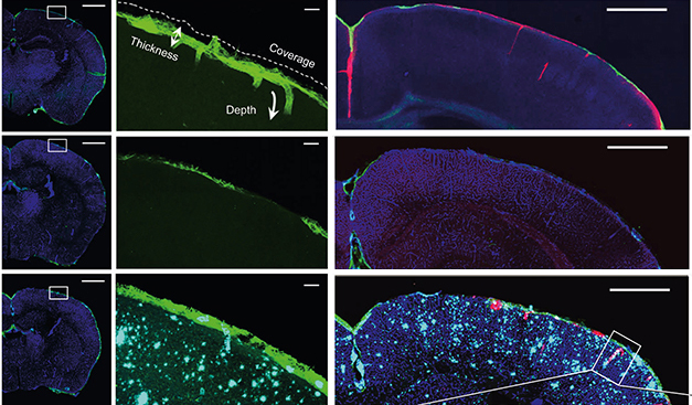
This surveillance may wane with age. Cortical surface tissue from 13-month-old mice had large areas without much pia or macrophages. This allowed more CSF to slip through (see image below). Mehta suggests this explains neuroimmune changes that occur along the brain surface with age. Tal Iram of Stanford University agreed. She was surprised by the intimate interaction of pia and immune cells. “Pial degeneration could be an upstream cause of many aging pathologies,” she told Alzforum.
The pia might also change in Alzheimer’s disease. In 13-month-old APP/PS1 mice, the pia was thick, trapping Aβ within its net. The fibrils blocked BSA injected into the cisterna magna from penetrating periarteriolar spaces (see image below). The APP/PS1 pia contained fewer macrophages than did same-age control pia. The authors hypothesized that either amyloid deposits in these spaces might disturb the integrity of pial cells and macrophage scaffolding sites, or that cell perturbations might encourage amyloid deposition within the pia itself. Intriguingly, the APP/PS1 macrophages took up more fluorescent BSA tracer than macrophages in wild-type mouse pia.

Young, Old, Amyloid. The pia of young mice (top) confines CSF (red) to the brain surface and perivascular spaces. The pia dissipates as the animals age (middle), allowing CSF to seep into the parenchyma. In APP/PS1 mice (bottom), the pia was abnormally thick, trapping pools of CSF and Aβ fibrils (turquoise). [Courtesy of Mestre et al., Nature Communications, 2022.]
“While it has been known that pia mater has fenestrations, the different patterns of perforated pia around arteries are novel, and may yield information on the pattern of dilated perivascular spaces related to the failure of drainage of interstitial fluid and cerebral amyloid angiopathy,” wrote Roxana Carare, University of Southampton, U.K. (see full comment below).
Mehta said her team members are now analyzing postmortem brain tissue from older adults with Alzheimer’s. They have already found some similarities and some differences to what they saw in mice.—Chelsea Weidman Burke
References
Research Models Citations
Paper Citations
- Hannocks MJ, Pizzo ME, Huppert J, Deshpande T, Abbott NJ, Thorne RG, Sorokin L. Molecular characterization of perivascular drainage pathways in the murine brain. J Cereb Blood Flow Metab. 2018 Apr;38(4):669-686. Epub 2017 Dec 28 PubMed.
Further Reading
News
- Lymphatic Brain Drain Withers in Aging, Worsens Disease
- Brain Drain—“Glymphatic” Pathway Clears Aβ, Requires Water Channel
- Not Just Aβ—Glymphatic Flow Clears Tau, Too, Slowing Its Aggregation
- More Evidence for Meningeal B Cells
- Sans Microglia, Mice Develop CAA and Die Young
- As Mice Age, T Cells Traipse Around Their Meninges. Mayhem Ensues
Primary Papers
- Mestre H, Verma N, Greene TD, Lin LA, Ladron-de-Guevara A, Sweeney AM, Liu G, Thomas VK, Galloway CA, de Mesy Bentley KL, Nedergaard M, Mehta RI. Periarteriolar spaces modulate cerebrospinal fluid transport into brain and demonstrate altered morphology in aging and Alzheimer's disease. Nat Commun. 2022 Jul 6;13(1):3897. PubMed.
Annotate
To make an annotation you must Login or Register.

Comments
University of Southampton School of Medicine
This paper provides new tools for a better understanding of the periarteriolar compartment. One problem that has held back the study of leptomeninges has been the lack of specific markers for leptomeninges. High-resolution three-dimensional microscopy, combined with the discovery of the ERTR7 marker, has shown the ultrastructure of the leptomeningeal coating of the arteries.
While it has been known that pia mater has fenestrations (Alcolado et al., 1988), the different patterns of perforated pia around arteries shown here are novel. They may yield information on the pattern of dilated perivascular spaces related to the failure of drainage of interstitial fluid and cerebral amyloid angiopathy.
It will be interesting to relate the findings in this study with the functions of pia mater as a regulatory interface between CSF and the cerebral parenchyma.
References:
Alcolado R, Weller RO, Parrish EP, Garrod D. The cranial arachnoid and pia mater in man: anatomical and ultrastructural observations. Neuropathol Appl Neurobiol. 1988 Jan-Feb;14(1):1-17. PubMed.
Make a Comment
To make a comment you must login or register.