With Age, Slower Protein Turnover May Predispose to Neurodegeneration
Quick Links
Problematic proteins are at the heart of neurodegenerative diseases, suggesting that as the brain ages, protein dynamics go off the rails. Now, a study published May 20 in Science Advances uncovered widespread evidence of unhinged proteostasis in older brains. In the most comprehensive analysis of its kind, researchers led by Anja Schneider of the German Center for Neurodegenerative Diseases, Bonn, and Eugenio Fornasiero of University Medical Center Göttingen, both in Germany, measured the half-lives of more than 3,500 proteins in the mouse brain. They found an average increase of 20 percent with age. Notably, lifespans of neurodegenerative disease-related proteins, e.g., APP, SORL1, cathepsin D, grew the most. Conversely, the half-lives of mitochondrial proteins shortened. Overall, turnover of aggregation-prone, heavily glycosylated, highly disordered, or heavy proteins that take a lot of energy to build slowed the most. The findings imply that, with age, changes in protein production, degradation, and aggregation fundamentally alter proteostasis in the aging brain and may be a prelude to neurodegeneration.
- Scientists measured the half-life of more than 3,500 proteins in mouse brain.
- Protein lifetime extended by 20 percent in brains of old mice.
- Neurodegenerative disease-related proteins among the most persistent.
- Mitochondrial proteins show opposite trend.
“These findings shed light on several mechanisms that regulate protein lifetime and stability in the brain [and] that may underlie shared defects in proteostasis observed across neurodegenerative diseases,” wrote Nicholas Seyfried of Emory University in Atlanta.
As the name implies, age-related proteinopathies are caused by malfunctions in protein homeostasis. What causes proteins in the brain to misbehave as the years roll by? To answer this question, scientists have measured the abundance of proteins in the brain. Such analyses have revealed only minor shifts with age, mostly in inflammatory proteins or proteasome and ribosome stoichiometry (Walther and Mann, 2011; Ori et al., 2015; Kelmer Sacramento et al., 2020).
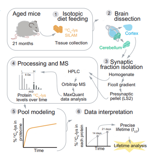
Another way to gauge proteostasis is to measure how long proteins last. “Their lifetimes indicate how fast proteins are replaced,” Schneider and Fornasiero wrote to Alzforum. “In practice, this is a measure of how ‘dynamic’ each protein is.” The authors reasoned that shifts in protein turnover might be chief among age-related changes that lead to neurodegenerative disease. Comprehensive studies of protein dynamics in the mammalian brain were lacking, so the scientists developed a technique to measure half-lives—aka lifetimes—of thousands of proteins in the mouse brain at once. In an in vivo adaptation of the stable isotope labeling with amino acids in cell culture (SILAC) technique, called “SILAM,” they fed mice a diet rich in 13C6-lysine and, after allowing this stable isotope to weave into newly translated proteins, the scientists then identified proteins from throughout the body via mass spectrometry, and inferred their half-lives (see image below).
Previously, Fornasiero and colleagues had used SILAM to measure the half-lives of some 3,500 proteins in the brains of 5-month-old mice, which are considered young adults (Fornasiero et al., 2018).

Analysis of a Lifetime. To measure the half-life of proteins in the brains of mice, scientists fed them a diet spiked with 13C6-lysine for different periods of time. They then took tissue from different brain regions and cellular fractions, and identified proteins via mass spectroscopy. Each protein’s half-life was determined by ratio of 13C6-to 12C6-lysine. [Courtesy of Kluever et al., Science Advances, 2022.]
For the current study, co-first authors Verena Kluever, Belisa Russo, Sunit Mandad, and colleagues applied the same type of analysis to six 21-month-old mice. The average half-life, which the scientists dubbed “lifetime,” of proteins in the aged cortex and cerebellum clocked in at 11.41 days, roughly 20 percent longer than the average protein sticks around in the brain of a 5-month-old whippersnapper. Fractionating synapses, the scientists found that these proteins tended to “live” 20 percent longer than proteins in the rest of the cellular homogenate. This slower turnover of synaptic proteins was true of both young and old brains.

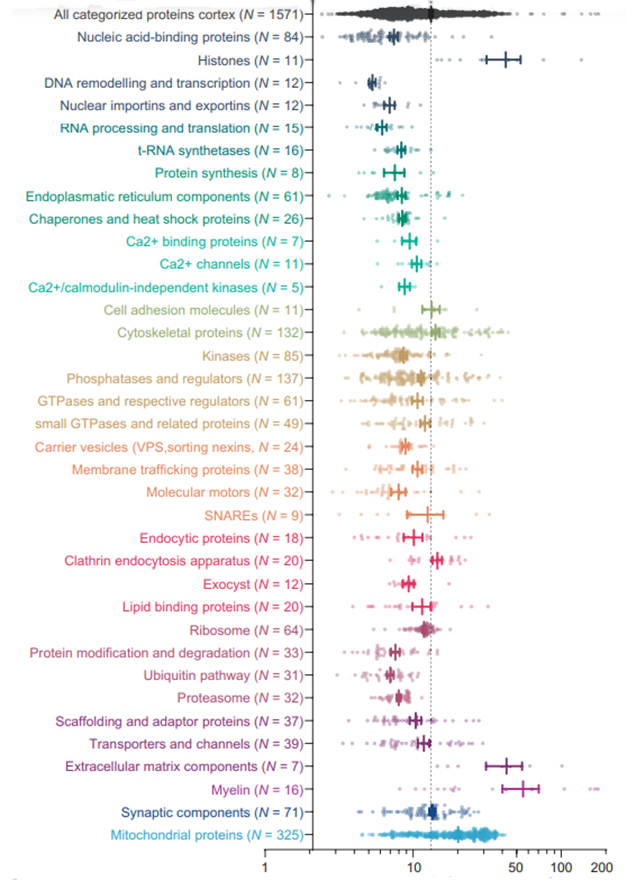
Spectrum of Lifetimes. Protein half-lives within the cortices of 21-month-old mice tracked with function. Transcriptional, translational, and cell-signaling proteins turn over sooner than do structural and housekeeping ones. [Courtesy of Kluever et al., Science Advances, 2022]
Did age alter the staying time of some proteins more than others? To address this, the researchers compared young versus old mice. They picked the 50 proteins that showed either the greatest extension or shortening with age as relatively longer lived (rLL) or relatively shorter lived (rSL), respectively. Surprisingly, proteins tied to neurodegenerative disease crowded into the rLL group. They included amyloid precursor protein (APP), sortilin-related receptor (SORL1), the lysosomal proteolytic enzyme cathepsin D (CTSD), prosaposin (PSAP), ferritin heavy chain 1 (FTH1), the neuroprotective carboxypeptidase E (CPE), and calsyntenin 1, which helps transport APP along axons. Many of these proteins are known to play neuroprotective roles, and their loss of function is associated with disease.
The longest-lasting proteins in both young and old mouse brain included those that provide structural integrity, such as histones, extracellular matrix proteins, and myelin components. Proteins with the most fleeting “lives” were those involved in dynamic processes, such as transcription, mRNA processing, and translation. Among neurodegenerative disease suspects, the researchers found wide variability in lifetimes that tended to correspond to function. For example, tau had a half-life of 19 days, which is similar to other proteins that bind to microtubules. At just 3.5 and 2.8 days, respectively, APP and SORL1 had shorter half-lives.
Why these proteins stay around longer in the aging brain is unclear, though the scientists are investigating the idea that their turnover becomes less efficient.
Though among the longest-lived in the brain proteome, mitochondrial proteins were prominent among the top rSLs, i.e., they turned over faster in older mice. The authors do not know why that is, but speculated that it could reflect increased turnover of the organelles, perhaps in an attempt to keep up with increased energy demands in the aging brain, and could lead to mitochondrial dysfunction.
The shortening half-life of mitochondrial proteins with age falls in line with another broad phenomenon the researchers uncovered: a compression of the range of protein lifespans. In other words, they found that with age, the proteins at the extremes of the lifespan spectrum moved closer to the average protein lifetime, i.e., half-lives of longer-lived proteins shortened while those of short-lived proteins lengthened. This reversion to a mean could reflect weakening homeostatic regulation, the authors speculated, which would preferentially affect the shortest- or longest-lived proteins that are under the tightest proteostatic regulation.
Interestingly, mitochondrial proteins had shortened half-lives with age whether they resided within synaptic fractions or in the rest of the cell, suggesting that age-related changes in mitochondrial turnover are not specific to synapses. In contrast, the lifetime of α-synuclein was extended only within the synaptic fraction, suggesting that age-related proteostatic changes involving this protein occur preferentially in the synapse.
What biochemical features might associate with lengthening or shortening of protein half-life? Highly aggregation-prone proteins and those with low-complexity domains tended to persist longer in the aged brain. Both are attributes of tau, TDP-43, and FUS. Heavily glycosylated proteins also stuck around longer in old brains, as did proteins flush with negatively charged amino acids. Interestingly, those more acidic proteins tend to be preferentially degraded in the lysosome, suggesting that they may linger longer as lysosomal function wanes with age. The authors also found that larger proteins, or those with many energetically expensive amino acids such as arginine, asparagine, or cysteine, live longer in the aged brain.
To Schneider and Fornasiero, this suggests that triage for turnover follows the “logic of proteomic cost minimization.” In other words, cells attempt to save energy as demands rise and metabolism flags with age. “This is an exciting finding since it might be one of those pre-pathological alterations occurring during brain aging,” they wrote.
This type of metabolic deficit could be amenable to lifestyle interventions such as diet. Some research suggests that caloric restriction may extend lifespan, in part by improving metabolism in the brain (Jul 2009 news; Sep 2013 news; Jan 2017 news).
In a comment to Alzforum, Kaspar Kepp of the Technical University of Denmark in Kongens Lyngby highlighted the findings linking lifetimes of disease-related and energetically costly proteins with aging. “If these relationships are causal in humans, it could explain why protein misfolding becomes pathogenic specifically in neurons, some of the most energy-demanding cells, and why this happens mainly at old age,” he wrote.
Nico Dantuma of the Karolinska Institute in Stockholm was intrigued by the idea that the aging brain preserves complex proteins to reduce energy expenditure, though he noted that it would be difficult to explain the molecular mechanisms involved in such selective stabilization. He wrote that hanging onto old, potentially malfunctioning proteins could have negative consequences, especially if the lingering proteins form insoluble aggregates. “This raises the question if the increased propensity of these ‘expensive,’ often large, proteins to aggregate could explain the more pronounced increase in their half-lives, since they may be harder to clear once they have aggregated,” he wrote.—Jessica Shugart
References
News Citations
- The Picture of Health? Aging Better—On Fewer Calories
- Brain-Specific Sirtuin Expression Delays Aging in Mice
- Consensus Reached: Dieting Monkeys Survive Longer
Paper Citations
- Walther DM, Mann M. Accurate quantification of more than 4000 mouse tissue proteins reveals minimal proteome changes during aging. Mol Cell Proteomics. 2011 Feb;10(2):M110.004523. Epub 2010 Nov 3 PubMed.
- Ori A, Toyama BH, Harris MS, Bock T, Iskar M, Bork P, Ingolia NT, Hetzer MW, Beck M. Integrated Transcriptome and Proteome Analyses Reveal Organ-Specific Proteome Deterioration in Old Rats. Cell Syst. 2015 Sep 23;1(3):224-37. Epub 2015 Sep 17 PubMed.
- Kelmer Sacramento E, Kirkpatrick JM, Mazzetto M, Baumgart M, Bartolome A, Di Sanzo S, Caterino C, Sanguanini M, Papaevgeniou N, Lefaki M, Childs D, Bagnoli S, Terzibasi Tozzini E, Di Fraia D, Romanov N, Sudmant PH, Huber W, Chondrogianni N, Vendruscolo M, Cellerino A, Ori A. Reduced proteasome activity in the aging brain results in ribosome stoichiometry loss and aggregation. Mol Syst Biol. 2020 Jun;16(6):e9596. PubMed.
- Fornasiero EF, Mandad S, Wildhagen H, Alevra M, Rammner B, Keihani S, Opazo F, Urban I, Ischebeck T, Sakib MS, Fard MK, Kirli K, Centeno TP, Vidal RO, Rahman RU, Benito E, Fischer A, Dennerlein S, Rehling P, Feussner I, Bonn S, Simons M, Urlaub H, Rizzoli SO. Precisely measured protein lifetimes in the mouse brain reveal differences across tissues and subcellular fractions. Nat Commun. 2018 Oct 12;9(1):4230. PubMed.
Further Reading
No Available Further Reading
Primary Papers
- Kluever V, Russo B, Mandad S, Kumar NH, Alevra M, Ori A, Rizzoli SO, Urlaub H, Schneider A, Fornasiero EF. Protein lifetimes in aged brains reveal a proteostatic adaptation linking physiological aging to neurodegeneration. Sci Adv. 2022 May 20;8(20):eabn4437. PubMed.
Annotate
To make an annotation you must Login or Register.

Comments
Emory University
In this interesting and well-conducted study, the authors measured the half-life or “lifetime” of brain proteins in mice using in-vivo metabolic labeling of amino acids and mass spectrometry. They found that in aged (21-month-old) compared to young brains (5-month-old), protein lifetimes were increased on average by 20 percent.
Interestingly, they note that many of the relatively longer-lived (rLL) proteins had key roles in neurodegenerative diseases including amyloid precursor protein (APP), sortilin-related receptor (SORL1), and the lysosomal proteolytic enzyme cathepsin D. Furthermore, the lifetime of α-synuclein was only seen increased in synaptic fractions suggesting that unique mechanisms control synuclein turnover in the synapse with age.
Alternatively, mitochondrial proteins were found to be relatively short-lived (rSL) with age, which may be associated with hypometabolic phenotypes with aging. The authors correlated protein lifetime with proteins previously identified in aggregate-rich fractions and saw a positive association with longer lived proteins being more prone to aggregate.
Another interesting finding is that the turnover for proteins with especially energetically expensive amino acid residues (e.g., cysteine, aspartate, asparagine) were significantly decreased in the aged brain. By extension, they show that, with age, rSL proteins become longer-lived and in turn rLL proteins become shorter-lived, suggesting that the brain gets “frugal” when it comes to protein synthesis over time.
Overall, these findings shed light on several mechanisms that regulate protein lifetime and stability in brain that may underlie shared defects in proteostasis observed across neurodegenerative diseases.
Technical University of Denmark
This collaboration of German labs is a major contribution to our field. Protein-misfolding-related neurodegenerative diseases are assumed to be caused by misfolded protein aggregates, or oligomers being toxic by some direct molecular mechanism of action, e.g., seeding other proteins to misfold or disrupting cell membranes. These aggregates and oligomers have thus been targeted in trials, notably via antibodies. How this fits with the major risk factor—aging—and why it is a particular problem to neurons has been much debated, as has the lack of success of clinical strategies.
While association is not causation and the new study used mice for necessary practical reasons, the study discovered consistent quantitative proteomic associations, with thousands of data points compared, between lifetimes of disease-related proteins and aging, and biosynthetic energy costs, with more expensive proteins being more preserved during aging.
The relationships provide the strongest indication so far that the underlying disease cause may perhaps not be a specific molecular toxic mode of action per se, but rather the indirect impact these proteins have on neuronal energy budgets.
If these relationships are causal in humans, it could explain why protein misfolding becomes pathogenic specifically in neurons, some of the most energy-demanding cells, and why this happens mainly at high age. It also points to new therapies. Antibodies may be too simplistic; rather, targeting the actual turnover of the oligomers and the associated metabolic consequences may be the avenues of research moving forward.
Karolinska Institutet
Using a clever mass-spectrometry method for determining protein turnover in vivo, the authors studied the effect of aging on protein lifetimes in the brains of mice. There were plenty of good reasons for having a closer look at protein turnover in the aged brain, as there is a large body of evidence indicating that protein synthesis as well as the major systems for protein degradation—the ubiquitin-proteasome system and autophagy—are dysregulated in the aged brain. This is likely to cause changes in protein homeostasis, i.e., proteostasis, which may explain the variety of age-related neurodegenerative diseases that are marked by the accumulation of protein aggregates in affected neurons.
An intriguing finding from this study is the observation that protein lifetimes on average were increased by 20 percent in aged brains, supporting the model that major changes in proteostasis occur in brains during aging. The data became even more revealing when the authors zoomed in on the effect of specific groups of proteins. Proteins that had a relatively longer half-life in the aged brain were typically involved in stress responses, autophagy, lysosome function, and other processes linked to neurodegeneration. Relatively long-lived proteins also had a slight tendency to be more disordered, suggesting that these proteins may have an increased propensity to aggregate. Interestingly, several proteins functionally linked to amyloid protein precursor (APP) trafficking and processing were among those relatively long-lived proteins.
On the other hand, an overrepresentation of mitochondrial proteins was found in the population of relatively short-lived proteins in the aged brains. This may reflect mitochondrial dysfunction, another phenomenon linked to age-related neurodegenerative disorders.
A fascinating but somewhat puzzling observation is that there seems to be a positive correlation between the bioenergetic cost of producing a protein and its lifetime in the aged brain, as “expensive” proteins had more extended lifetimes than “cheap” proteins in the aged brains. Admittedly, it may be beneficial for cells to make optimal use of these “expensive” proteins from an energy expenditure point of view, but the molecular mechanism that would be responsible for the selective stabilization of these proteins may be harder to explain. Aged brains hanging on these proteins longer than would have been the case in younger brains may sound attractive, but may also increase the risk of old, malfunctioning proteins forming insoluble aggregates or otherwise disturbing cellular functions.
This also raises the question if an increased propensity to aggregate of these “expensive”, often large, proteins could be a cause for the more pronounced increase in their half-lives, as they may be harder to clear once aggregated. As this line of reasoning exemplifies, any attempt at explaining these data with underlying molecular mechanisms is, at this point, largely a matter of conjecture. Insights into causality and identification of confounding factors will be important, but would require additional experimentation on underlying molecular mechanisms.
This study undoubtedly provides the field with a wealth of important information that can form steppingstones for better understanding the effect of aging on proteostasis in the brain as well as its link to neurodegenerative disease, but additional mechanistic studies will be critical to learn how and why these changes in protein turnover happen. That will be the next challenge.
University of Kansas
This very well-done and very interesting proteomics study provides a fundamental snapshot of how protein biology changes in the aging brain, or at least in this case in the aging mouse brain. The unbiased analysis reveals there are proteins and protein modules whose existence time increases, and proteins and protein modules whose existence time decreases. Perhaps this by itself is not surprising, but what is clearly interesting is the nature of the modules in which protein longevity increases versus decreases.
The authors broadly note two classes of long-lived proteins: (1) those that may lead to adaptive resilience under stress, and (2) those that associate for one reason or another with neurodegenerative diseases. This raises the question of whether some of the latter associate with neurodegenerative diseases because they are driving aging or disease, or are responding to aging or disease.
The data also reveal that proteins that become relatively short-lived are enriched for mitochondrial proteins. This raises the question of whether the changes in the mitochondrial proteins drive age-related changes in mitochondrial function or are driven by age-related changes in mitochondrial function. Finally, it appears that proteins with higher biosynthetic costs end up hanging around longer, which could reflect an attempt by neurons to conserve energy.
I thought the authors did a good job in considering the implications of their findings. One really can’t tell the extent to which the proteostasis changes contribute to aging, are a consequence of aging, or both. Regardless, this study leaves us with a lot to think about. We may not know the answer to these questions at this time, but I am struck by the fact that these data were generated in aging mice, that changing mitochondrial biology is a hallmark of aging, that strategic adaptations to energy stress are implicated, and the data provide links to neurodegenerative diseases. All this is arguably consistent with the view that mitochondria potentially play a relatively upstream role in an age-related disease such as AD.
Make a Comment
To make a comment you must login or register.