That Retrotransposon in TMEM106b: Friend or Foe?
Quick Links
Ever since variants in the gene for TMEM106b were tied to frontotemporal dementia, Alzheimer’s, and other neurodegenerative diseases, this endolysosomal protein has been a head-scratcher for scientists. Its ability to surprise is exemplified by the discovery of fibrils spun of the protein—but there’s much more. According to findings presented at the Alzheimer’s Association International Conference, held July 27 to August 1 in Philadelphia, an AluYb8 retrotransposon is situated within its 3ˈ untranslated region. Geneticists spotted it by scouring long-read sequences in search of structural variants around the gene. The twist? This retroviral relic was found sitting in the risk-boosting variant. Cognitively resilient centenarians tended to carry the protective, transposon-free version.
- An AluYb8 transposon was found within the 3’UTR of a common risk allele of TMEM106b.
- Cognitively healthy centenarians tend to carry a protective TMEM106b variant without the retrotransposon.
- Populations of African ancestry harbor alternative haplotypes of TMEM106b.
Importantly, this wee element represents the tip of an iceberg. The scientists found myriad other repeat sequences and expansions stationed near the gene. Moreover, different combinations of structural variants occur in people of African ancestry.
How this mounting pile of genomic variations affects TMEM106b’s expression, proper function, and dysregulation is a new area ripe for study. In Philadelphia, Henne Holstege of Amsterdam University Medical Center proposed a potential mechanism linking age-related demethylation, TDP-43 aggregation, and TMEM106b’s awakening transposon with neurodegenerative pathogenesis.
TMEM106b burst onto the neurodegeneration scene almost 15 years ago, when geneticists identified common variants in the gene that either increased or decreased a person’s risk of FTD and ALS (Feb 2010 news; Aug 2012 news). Later, TMEM106b’s sphere of influence expanded to AD and other neurodegenerative diseases (Sep 2021 news).
What might it do? TMEM106b knockout mice displayed endolysosomal dysfunction, which was worse in mice that also lacked progranulin (Sep 2020 news). Just when scientists thought they were getting a handle on TMEM106b’s modus operandi, structural biologists joined the fray and unveiled TMEM106b fibrils lurking in brain samples from people with neurodegenerative diseases and in many cognitively normal people older than 50 (Apr 2022 news). Subsequently it appeared that TMEM106b’s propensity to fibrillize goads TDP-43 pathology, which itself underlies many cases of FTD/ALS as well as limbic predominant TDP-43 encephalopathy (LATE) (Jan 2024 news). Exactly how genetic variation at the TMEM106b locus relates to its fibrillization and how that might tempt other proteopathic culprits to follow suit remain big puzzles in the field.
For Holstege’s team, protective variants in TMEM106b surfaced as top genetic discriminators of sharp centenarians. Holstege heads the 100-plus Study, which tracks comprehensive molecular, genetic, neuropathological, and health attributes of people who have managed to remain cognitively healthy into their 11th decade of life (Holstege et al., 2018). The study includes some 480 participants. They sit for annual testing, and many donate their brains after they pass on.
So far, Holstege has found that at autopsy, these centenarians had less brain Aβ, tau, and TDP-43 pathology than did people who died at a younger age with AD. Crucially, they also enjoyed remarkable cognitive resilience in the face of whatever proteopathy they did have until shortly before death (Zhang et al., 2022; Zhang et al., 2023).

Endolysosomal Endurance. Centenarians sported protective variants, or lacked risky ones, in three genes encoding proteins needed in endolysosomes: progranulin, sortilin, and TMEM106b. [Courtesy of Holler et al., eNeuro, 2017.]
How did they fend off these age-related scourges? In Philadelphia, Holstege presented results from a genetic analysis. First, postdoc Niccoló Tesi asked if the centenarians carried protective variants versus variants tied to increased AD risk. For 85 percent of the AD GWAS hits Tesi looked at, the cognitively healthy centenarians had a higher frequency of protective and lower frequency of risk alleles, relative to middle-aged people with AD or their age-matched controls. As such, the centenarians also had lower polygenic risk scores, and were likelier to carry the protective ApoE2 allele and unlikelier to carry ApoE4. Notably, protective variants in a trio of endolysosomal genes—SORT1, GRN, and, you guessed it, TMEM106b—were among those most strongly overrepresented in the centenarians (Tesi et al., 2024).
Next, Holstege noted that several risk TMEM106b SNPs had popped up in GWAS for AD, FTD, and ALS, including the coding variant T185S. They all corresponded to the same haplotype. Yet single-nucleotide polymorphisms are only one part of the picture of genetic variation. Also important are structural variants, such as short and long tandem repeats, transposable elements, and methylated CpGs, the latter of which regulate gene expression.
Only long-read sequencing can detect these structural changes. Postdoc Alex Salazar performed this new type of analysis on the genomes of 250 people with AD and 251 resilient centenarians. Lo and behold, he found a 317-bp AluYb8 retrotransposon lurking within the 3ˈUTR of people who carried the SNPs corresponding to the risk-raising allele, but not the protective one. The allele infiltrated by the transposon was enriched in people with AD; the protective one in centenarians (image below).

Retroviral Interloper. An AluYb8 retrotransposon (pink) was found lurking within the risk-boosting haplotype of TMEM106b (top), not within the protective variant (bottom). [Courtesy of Henne Holstege, Amsterdam University Medical Center.]
How might this mobile element affect TMEM106b’s expression or function? In Philadelphia, Holstege told the audience that Alu elements are retrotransposons that make up a whopping 11 percent of the human genome. They use a copy-and-paste strategy—i.e., reverse transcription and insertion, respectively—to propagate themselves across the genome. This frivolous behavior can disrupt expression and regulation of nearby genes, such as TMEM106b. In response, methylation of surrounding CpG sequences has evolved to ground the jumping genes. As methylation is known to wane with age, Holstege proposed that the Alu element might become increasingly active, potentially messing with TMEM106b expression among those who carry the risk allele.
As evidence that these mechanisms might be at play, Holstege reported at AAIC that 52 CpG sites in and around the TMEM106b gene were significantly likelier to be methylated in people carrying the risk haplotype than in people carrying the transposon-free one. Furthermore, the risk haplotype includes 19 unique CpG sites, hinting that the genome evolved to put the kibosh on the nearby Alu element, Holstege said.
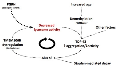
Connecting the dots between TMEM106b and TDP-43, Holstege pointed out that TDP-43 has been reported to bind and suppress Alu elements (Morera et al., 2019). Moreover, TDP-43 itself becomes increasingly demethylated with age in the brain, hampering TDP-43’s normal autoregulation (Koike, 2024). The resulting glut of TDP-43 could then lead to its accumulation and aggregation, ultimately sequestering it from performing its functions—Alu suppression among them.

Demethylate, Degenerate? With advancing age, TMEM106b demethylation might stir up the AluYb8 retrotransposon. TARDBP demethylation and TDP-43 aggregation might also awaken the transposon, as physiological TDP-43 suppresses it. [Courtesy of Henne Holstege, Amsterdam University Medical Center.]
Holstege noted that TDP-43 pathology is seen in people with AD, FTD/ALS, and LATE, but rarely in centenarians. Therefore, she proposed that age-related demethylation may lift the lid on TMEM106b’s transposon in two ways: one, via demethylating TMEM106b itself, and two, by demethylating the Alu silencer, TDP-43 (image below). The findings are posted on medRxiv (Salazar et al., 2023).
Just how an activated AluYb8 element in TMEM106b’s 3ˈUTR affects the protein’s expression, much less fibrillization, remains unclear, Salazar told Alzforum. Two other groups found the same Alu element in the risk allele. One, led by Michael Koob of the University of Minnesota in Minneapolis, discovered the insertion when sequencing the TMEM106b region in preparation to make mouse models (Rodney et al., 2024). The other, led by Michael Greicius of Stanford University, reported that carriers of the transposon variant had more TMEM106b in their plasma and CSF (Chemparathy et al., 2023). “The mechanism by which the insertion may impact TMEM106B levels remains uncertain,” they wrote, proposing that changes in the 3ˈUTR could affect protein binding, translation efficiency, even subcellular localization of the mRNA. “Identifying a clear-cut mechanism linking the insertion to increased TMEM106B protein levels is still required to confirm that this is the causal variant at the locus,” they added.
Beyond Alu
But wait, even that is not all. In Philadelphia, Salazar reported that the Alu element was but one of many structural variants riddling the TMEM106b region. “There’s a lot more going on in this region than previously understood,” he told the audience. Case in point, just upstream of TMEM106b’s transcriptional start site, Salazar discovered a large, 4.6-kilobase stretch housing a slew of transposable elements. In a lone centenarian, this transposon stretch was missing. Salazar also unearthed a string of tandem AC repeats located 3ˈ of the Alu element. This sequence was multi-allelic, ranging from 35 to 80 bp in length. In all, Salazar spotted more than 60 structural variants located in or around the TMEM106b gene.
How do these genomic hijinks relate to the single-nucleotide variants that tracked with disease risk in GWAS? Salazar examined all of these variants at once. He generated more than 1,000 “haplotype fingerprints,” one for each copy of the locus carried by all participants in the 100-plus cohort. In this way, he identified a new protective haplotype. It lacks the Alu element but has different combinations of other structural variants relative to the original protective allele. This variant combo had double the protective power of the original, he reported.
This diversity of variants occurs in a homogenous population, i.e., people of European ancestry living in the Netherlands. How might this TMEM106b locus look in other ancestries? To find out, Salazar integrated whole genome sequencing data from the Human Pangenome Reference Consortium. Among people of African ancestry, he discovered two novel haplotypes. One included the SNPs corresponding to the risk-promoting allele but its 3ˈUTR matched the protective haplotype devoid of the Alu element. The other was the opposite, suggesting recombination events had produced the hybrid alleles. How these new combinations influence disease risk has yet to be investigated. In line with TMEM106b’s penchant for complexity, the researchers, even since AAIC, have unearthed even more unique haplotypes. They will describe them in an upcoming preprint.
Finally, in a separate AAIC talk, Tesi expanded the scope of the analysis beyond the tiny neighborhood of TMEM106b. Wondering if structural variants positioned near GWAS hits might be the actual drivers of disease, Tesi used long-read sequencing to explore the interplay between large such variants and AD risk SNPs in the 100-plus cohort. He identified 27,404 structural variants averaging 763bp in length, including more than 7,600 transposable elements and 17,500 tandem repeats. Among 85 GWAS hits, 37 were closely linked with a structural variant. Some, including ADAM10, SLC2A4RG, and CD2AP, were tied to more than one. For three genes—APOC1, SPI1, and ABCA7—the researchers found a significant association between the structural variant and AD risk within their cohort.
All told, the preliminary findings hint that structural variants might contribute to AD risk imparted by those genes, Tesi said. Pulling the most important pathways out of this genomic melee will take some time.—Jessica Shugart
References
News Citations
- Genetics of FTD: New Gene, PGRN Variety, and a Bit of FUS
- FTD Risk Factor Confirmed, Alters Progranulin Pathways
- From a Million Samples, GWAS Squeezes Out Seven New Alzheimer's Spots
- Nixing TMEM106b Fans the Flames of Progranulin Deficiency
- Surprise! TMEM106b Fibrils Found in Neurodegenerative Diseases
- In FTD, TMEM106b Fibrils Tip TDP-43 Dysfunction into Overdrive
Paper Citations
- Holstege H, Beker N, Dijkstra T, Pieterse K, Wemmenhove E, Schouten K, Thiessens L, Horsten D, Rechtuijt S, Sikkes S, van Poppel FW, Meijers-Heijboer H, Hulsman M, Scheltens P. The 100-plus Study of cognitively healthy centenarians: rationale, design and cohort description. Eur J Epidemiol. 2018 Dec;33(12):1229-1249. Epub 2018 Oct 25 PubMed.
- Zhang M, Ganz AB, Rohde S, Rozemuller AJ, Bank NB, Reinders MJ, Scheltens P, Hulsman M, Hoozemans JJ, Holstege H. Resilience and resistance to the accumulation of amyloid plaques and neurofibrillary tangles in centenarians: An age-continuous perspective. Alzheimers Dement. 2022 Dec 30; PubMed.
- Zhang M, Ganz AB, Rohde S, Lorenz L, Rozemuller AJ, van Vliet K, Graat M, Sikkes SA, Reinders MJ, Scheltens P, Hulsman M, Hoozemans JJ, Holstege H. The correlation between neuropathology levels and cognitive performance in centenarians. Alzheimers Dement. 2023 Apr 24; PubMed.
- Tesi N, van der Lee S, Hulsman M, van Schoor NM, Huisman M, Pijnenburg Y, van der Flier WM, Reinders M, Holstege H. Cognitively healthy centenarians are genetically protected against Alzheimer's disease. Alzheimers Dement. 2024 Jun;20(6):3864-3875. Epub 2024 Apr 18 PubMed.
- Morera AA, Ahmed NS, Schwartz JC. TDP-43 regulates transcription at protein-coding genes and Alu retrotransposons. Biochim Biophys Acta Gene Regul Mech. 2019 Oct;1862(10):194434. Epub 2019 Oct 23 PubMed.
- Koike Y. Molecular mechanisms linking loss of TDP-43 function to amyotrophic lateral sclerosis/frontotemporal dementia-related genes. Neurosci Res. 2024 May 8; PubMed.
- Salazar A, Tesi N, Knoop L, Pijnenburg Y, vanderLee S, Wijesekera S, Krizova J, Hiltunen M, Damme M, Petrucelli L, Reinders M, Hulsman M, Holstege H. An AluYb8 retrotransposon characterises a risk haplotype of TMEM106B associated in neurodegeneration. 2023 Oct 03 10.1101/2023.07.16.23292721 (version 3) medRxiv.
- Rodney A, Karanjeet K, Benzow K, Koob MD. A common Alu insertion in the 3'UTR of TMEM106B is associated with risk of dementia. Alzheimers Dement. 2024 Jul;20(7):5071-5077. Epub 2024 Jun 26 PubMed.
- Chemparathy A, Guen YL, Zeng Y, Gorzynski J, Jensen T, Yang C, Kasireddy N, Talozzi L, Belloy ME, Stewart I, Gitler AD, Wagner AD, Mormino E, Henderson VW, Wyss-Coray T, Ashley E, Cruchaga C, Greicius MD. A 3'UTR Insertion Is a Candidate Causal Variant at the TMEM106B Locus Associated with Increased Risk for FTLD-TDP. medRxiv. 2023 Nov 17; PubMed.
External Citations
Further Reading
Annotate
To make an annotation you must Login or Register.

Comments
No Available Comments
Make a Comment
To make a comment you must login or register.