Blood Tests of Phospho-Tau, Aβ42, Track With Brain Amyloid
Quick Links
While a suite of new CSF markers has entered a mature stage where they get validated with identical methods in large international cohorts (see previous CTAD story), much newer blood tests are catching up fast. Scientists are pushing the limits of detection of the core AD markers in plasma, and in the process are cracking open what used to be considered the single category of p-tau into a bewildering new spectrum of subspecies. At CTAD, Nicolas Barthélemy of Washington University, St. Louis, showed what this looked like when he presented fresh mass spectrometry data on different forms of plasma phospho-tau. Compared with immunoassays, which rely on antibodies trained against a priori designated phospho-epitopes, mass spectrometry gives an unbiased, and exquisitely specific, view of the landscape of phospho-tau isoforms in a given sample.
- Mass spectrometry picked up the lowest concentration of phospho-tau ever detected in plasma.
- P-tau-217 picked out people with brain amyloid even better than p-tau-181.
- A plasma test for Aβ42 highly correlated with amyloid PET, and seeks regulatory approval.
Previously, Barthélemy and colleagues put mass spec to use in CSF samples. At AAIC earlier this year, he wowed the crowd by reporting that, in samples collected from the DIAN cohort, CSF p-tau-T217 started rising in CSF just after Aβ started accumulating in the brain—fully 20 years prior to estimated symptom onset (Aug 2019 news). CSF p-tau-181—the epitope most commonly measured in biomarker immunoassays—trailed close behind, at 19 years prior to onset.
At CTAD, Barthélemy presented his mass spectrometry data on plasma tau. This project posed unique challenges. For one, at less than 1 pg/mL, the concentration of p-tau in plasma is vanishingly low, while the complexity of other proteins in this bodily fluid is exorbitant, making p-tau detection there akin to searching for a needle in a haystack. For another, while mass spectrometry is highly specific, it is less sensitive than immunoassays. Compared with the 0.2 mL required to detect p-tau-T181 using the most sensitive immunoassay available, Barthélemy needed 20 mL of plasma to detect it via mass spec. That translates to a 40 mL blood draw, which is not feasible on a large clinical scale, researchers agree. For a typical blood panel in routine medical care, a phlebotomist draws 5–10 mL. This is why mass spec is particularly well suited for discovery research, but will need to become dramatically more sensitive for large-scale use or population screening.
It so happened that Barthélemy had access to large volumes of plasma that had been collected from 35 participants at the Knight Alzheimer’s Research Center during a stable isotope labeling kinetics (SILK) study that tracked the half-life of tau (Mar 2018 news). Twenty of them had tested negative for Aβ accumulation by CSF-Aβ42/40 and amyloid -PET, while 15 were positive. Among the latter, five were cognitively normal, eight were at the stage of MCI due to AD, and two had moderate AD dementia. As was the case in DIAN, too, only the amyloid-positive, cognitively impaired participants had significant accumulation of tangles as assessed by tau PET. In contrast, CSF measurements of p-tau-T181 via mass spec were elevated earlier, in people with amyloid plaques who were still cognitively normal.

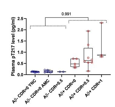
P-tauT217 in Plasma: Aβ in Brain. Plasma concentrations of p-tau-T217 were elevated in people harboring Aβ deposits in the brain. [Courtesy of Nicolas Barthélemy, Washington University, St. Louis.]
But what about their plasma? Similar to what Barthélemy observed in CSF, he found tau peptides of 14 different lengths, most of which were truncated at the C-terminus. However, of the 11 unique phosphorylated sites Barthélemy was able to detect in CSF, only four were detectable in plasma: p-tau-T181, p-tau-S202, p-tau-T205, and p-tau-T217. Levels of both p-tau-T181 and p-tau-T217 were significantly higher in plasma from amyloid-positive than -negative participants, including those who were cognitively normal. While p-tau-T181 was 74 percent higher, p-tau-T217 was up by a whopping 365 percent, an exponentially more robust difference than detectable with Aβ42/40 ratio tests. Moreover, p-tau-T217 in plasma correlated closely with p-tau-T217 concentration in CSF, suggesting that its blood level reflects processes in the brain.
In contrast, Barthélemy found that, for both plasma total tau and p-tau-S202, their concentration was about the same in people with and without brain amyloid. He believes p-tau-S202 gets released from cells outside the brain, in which tau is more highly phosphorylated on serine 202 than is tau in the CNS. A high background level of p-tau-S202 in the periphery would mask changes in p-tau-S202 contributed by the CNS. On the other hand, p-tau-181 and p-tau-217 are more specific to the CNS, allowing for detection of subtle differences in the plasma. As for p-tau-T205, Barthélemy only detected it in some plasma samples from the cohort, and its concentration was exceedingly low. He told Alzforum that this phospho-species is likely specific to the CNS, and exists at too low a concentration for consistent detection in plasma even with current mass spec methods.
Will plasma p-tau tests pick up other tauopathies, such as progressive supranuclear palsy or frontotemporal dementia? Barthélemy said that as of now, he has no access to cohorts with sufficient plasma to address this question. Just as is being done in AD, p-tau species that change in other tauopathies will need to be validated in CSF before digging for them in plasma.
Barthélemy acknowledged that the volume of plasma needed to detect p-tau by mass spectrometry precludes implementing the MS-based test on a large scale. MS assays are becoming more sensitive, however. SEABIRD, a St. Louis area study to learn how well MS-based tests pick up CNS Aβ too, is currently validating a protocol that requires only 4 mL of plasma.
For his part, Zetterberg and colleagues are using mass spec as a guide to help them develop sensitive immunoassays, especially for p-tau-T217. At CTAD, Zetterberg talked about measuring p-tau-T181 in plasma using the Simoa platform. In samples from the McGill Translational Biomarkers in Aging and Dementia (TRIAD) cohort, Zetterberg reported that p-tau-181 was two- to threefold higher in people with MCI or AD, compared with cognitively normal elderly people. Yes, fold. Not a few percent higher.
This agrees with findings reported by other groups using different antibodies and platforms, including researchers at Eli Lilly in Indianapolis, the Swedish BioFINDER group at Lund University, as well as researchers at the University of California, San Francisco (Mielke et al., 2018, and Aug 2019 conference news). Zetterberg noted that p-tau-T181 has been more extensively validated than p-tau-T217 as an AD biomarker, both in CSF and plasma, and therefore will likely be the first to garner approval as a bona fide AD screening test. If sensitive immunoassays for p-tau-217 are developed and validated in multiple cohorts, it could follow suit.
A test for plasma Aβ is already moving through the paces for regulatory approval, though it has to squeak by with measuring a less than 20 percent difference between groups. At CTAD, Tim West of C2N Diagnostics, a company formed 12 years ago by WashU researchers Randall Bateman and David Holtzman and LifeTech Research, Inc., reported fresh results from C2N’s mass spectrometry-based test for the plasma Aβ42/40 ratio.
Previously, Bateman had reported that among 158 participants in the Knight Alzheimer’s Disease Research Center, the plasma Aβ42/40 ratio correlated tightly with Aβ-PET (Aug 2019 conference news on Schindler et al., 2019). People with elevated plasma Aβ42/40 ratios whose amyloid PET scans were negative were 15 times more likely to be positive on a subsequent scan taken two to nine years later, suggesting the plasma test was predictive of amyloidosis in the near future.
At CTAD, West extended those findings to a more-diverse, less-standardized group of samples that are part of C2N’s clinical validation study. He analyzed 415 plasma samples collected from six independent biobank cohorts, in which blood was collected, handled, and stored in different ways, and Aβ positivity was independently determined using different methods (i.e., CSF versus PET, with a variety of tracers). Thirty-nine percent of the combined cohort harbored brain amyloid, according to these independent methods. Participants averaged 70 years of age, and among 309 people with ethnicity data, 23 percent were Hispanic. Forty percent carried at least one copy of ApoE4. West reported an 81 percent concordance between the Aβ42/40 plasma test and amyloid status. In a receiver operator curve analysis—which measures the probability that a given measure will predict a specific outcome—the results translated to probability of 0.86, which improved to 0.90 once the researchers factored in age and ApoE4 status.
Consistent with Bateman’s previous study, the plasma test produced more false positives than false negatives. West hypothesized that those false positives were not truly false, but rather a reflection of using a binary outcome to classify what is in reality a disease continuum. In other words, these “false positives” might lie just below the threshold of detection via PET. If this is the case, these people, too, would be expected to have a positive scan in the near future.
C2N is currently validating their ratio test in samples from participants in the PARIS study, who had amyloid PET scans as part of the Imaging Dementia—Evidence for Amyloid Scanning (IDEAS) study. Based on data collected from this sample set, C2N will seek approval for the test as an in vitro diagnostic.—Jessica Shugart
References
News Citations
- Move Over Aβ, CSF P-Tau Tells Us There’s Plaque in the Brain
- Isotope Labeling Links Tau Production to Aβ Burden
- Move Over CSF, P-Tau in Blood Also Tells Us There’s Plaque in the Brain
- Are Aβ Blood Tests Ready for Prime Time?
Paper Citations
- Mielke MM, Hagen CE, Xu J, Chai X, Vemuri P, Lowe VJ, Airey DC, Knopman DS, Roberts RO, Machulda MM, Jack CR Jr, Petersen RC, Dage JL. Plasma phospho-tau181 increases with Alzheimer's disease clinical severity and is associated with tau- and amyloid-positron emission tomography. Alzheimers Dement. 2018 Aug;14(8):989-997. Epub 2018 Apr 5 PubMed.
- Schindler SE, Bollinger JG, Ovod V, Mawuenyega KG, Li Y, Gordon BA, Holtzman DM, Morris JC, Benzinger TL, Xiong C, Fagan AM, Bateman RJ. High-precision plasma β-amyloid 42/40 predicts current and future brain amyloidosis. Neurology. 2019 Oct 22;93(17):e1647-e1659. Epub 2019 Aug 1 PubMed.
Other Citations
Further Reading
No Available Further Reading
Annotate
To make an annotation you must Login or Register.

Comments
No Available Comments
Make a Comment
To make a comment you must login or register.